
Device and methods for color corrected oct imaging endoscope/catheter to achieve high-resolution

Active Publication Date: 2016-08-11
THE JOHN HOPKINS UNIV SCHOOL OF MEDICINE
View PDF6 Cites 6 Cited by
  • Summary
  • Abstract
  • Description
  • Claims
  • Application Information

AI Technical Summary

Benefits of technology

The patent describes a method for improving the resolution of OCT imaging by using a special diffraction element in the imaging optics of the probe and reducing the focal shift of the light source. This results in better lateral resolution and minimizes distortion to the reflected light, allowing for optimal axial resolution. Overall, this method helps to achieve higher quality OCT images.

Problems solved by technology

Chromatic aberration in the OCT imaging optics will alter the backreflected spectrum from the target, resulting in the loss of OCT axial resolution.
The change in the backreflected spectrum from the sample could also result in the increase in the side lobes of the OCT imaging signal, which again will lead to the loss of OCT axial resolution.
But, such approaches are not cost effective or practical to be implemented in miniature OCT imaging probes.
For the endoscopic setting, due to the challenges such as management of chromatic aberration over a broadband spectral bandwidth, there are only few achromatic endoscopic setups.
The designs in those endoscopes are rather complicated and expensive, involving multi-element achromatic microlenses.

Method used


Image

  • Device and methods for color corrected oct imaging endoscope/catheter to achieve high-resolution
  • Device and methods for color corrected oct imaging endoscope/catheter to achieve high-resolution
  • Device and methods for color corrected oct imaging endoscope/catheter to achieve high-resolution

Examples


Embodiment Construction

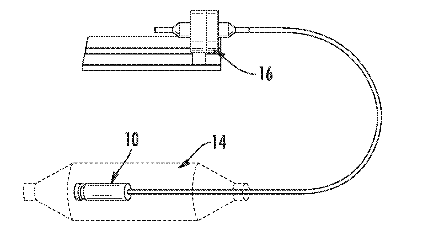
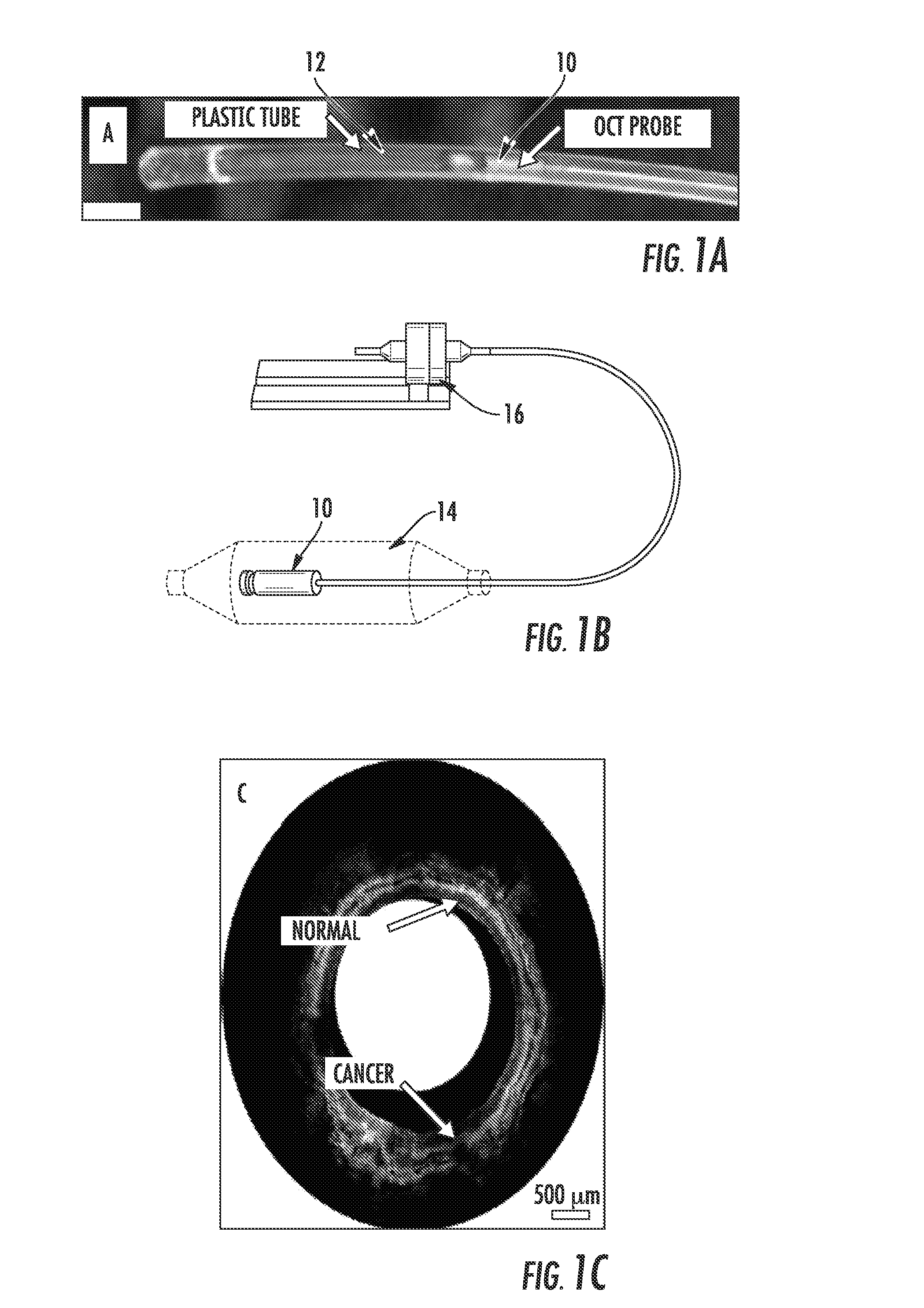
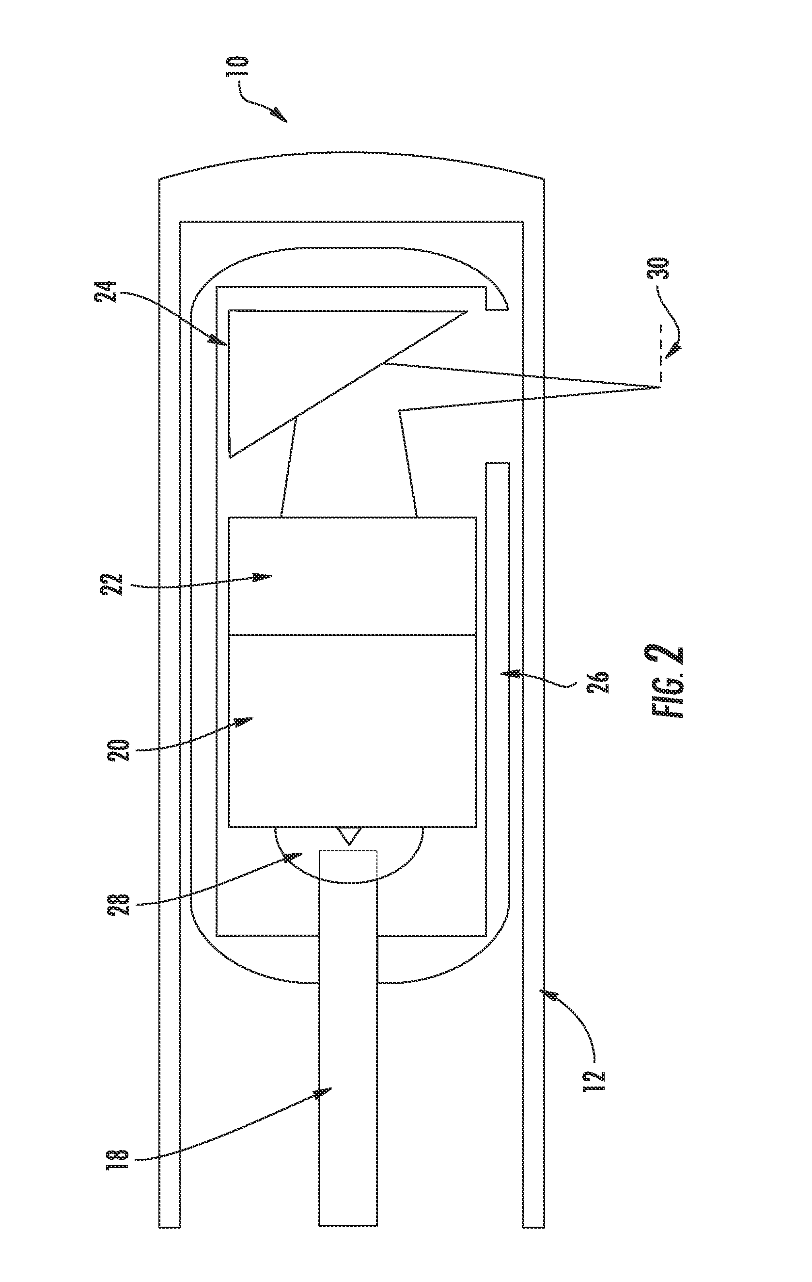
[0026]The presently disclosed subject matter now will be described more fully hereinafter with reference to the accompanying Drawings, in which some, but not all embodiments of the inventions are shown. Like numbers refer to like elements throughout. The presently disclosed subject matter may be embodied in many different forms and should not be construed as limited to the embodiments set forth herein; rather, these embodiments are provided so that this disclosure will satisfy applicable legal requirements. Indeed, many modifications and other embodiments of the presently disclosed subject matter set forth herein will come to mind to one skilled in the art to which the presently disclosed subject matter pertains having the benefit of the teachings presented in the foregoing descriptions and the associated Drawings. Therefore, it is to be understood that the presently disclosed subject matter is not to be limited to the specific embodiments disclosed and that modifications and other ...


PUM


Abstract

The present invention is directed to an achromatic endoscope which employs a diffractive microlens. Along with a broadband rotary joint and a custom 800 nm SD-OCT system, ultrahigh-resolution 3D volumetric imaging over a large area becomes possible. The diffractive microlens can be used directly with a GRIN lens, making the endoscope design simpler and cost effective. Preliminary ex vivo 3D intraluminal imaging was performed with the endoscope in conjunction with a home-built broadband rotary joint and a spectral-domain OCT system, demonstrating the performance of the diffractive endoscope. Considering the miniature OCT imaging probe is the required component for using the OCT technology in internal organs, the proposed approach will have a broad impact on endoscopic OCT imaging by improving OCT resolution in any applications that involve a miniature OCT probe, as intravascular OCT imaging, gastrointestinal (GI) tract imaging, airway imaging etc.

Description

CROSS REFERENCE TO RELATED APPLICATIONS[0001]This application claims the benefit of U.S. Provisional Patent Application No. 61 / 878,885 filed on Sep. 17, 2013 and to U.S. Provisional Patent Application No. 61 / 948,826 filed on Mar. 6, 2014, both of which are incorporated by reference, herein, in their entirety.GOVERNMENT SUPPORT[0002]This invention was made with government support under R01EB007636 awarded by the National Institutes of Health and the National Institute of Biomedical Imaging and Bioengineering and R01CA153023 awarded by the National Institutes of Health and the National Cancer Institute. The government has certain rights in the invention.FIELD OF THE INVENTION[0003]The present invention relates generally to medical imaging. More particularly, the present invention relates to a device and method for color corrected OCT imaging.BACKGROUND OF THE INVENTION[0004]OCT is a noninvasive, high-resolution optical imaging technology capable of real-time imaging of tissue microana...

Claims


Application Information

Patent Timeline
IPC IPC(8): A61B1/00A61B5/00
CPCA61B1/00096A61B1/00172A61B5/0084A61B5/0066A61B1/002
Inventor LI, XINGDEZHANG, ANQIXI, JIEFENG
Owner THE JOHN HOPKINS UNIV SCHOOL OF MEDICINE
Learn More