Tracking-based 3D model enhancement

a 3d model and tracking technology, applied in the field of medical imaging and positioning systems, can solve the problems of loss of the beneficial effect of the contrast agent highlighting the anatomical structure, the inability to capture an anatomy image, and the inability to capture images simultaneously showing medical devices and images, etc., to achieve the effect of enhancing the data acquired and enhancing the three-dimensional (3d) reconstruction

Active Publication Date: 2016-08-18
ST JUDE MEDICAL INT HLDG SARL
View PDF37 Cites 18 Cited by
  • Summary
  • Abstract
  • Description
  • Claims
  • Application Information

AI Technical Summary

Problems solved by technology

Oftentimes, though, it is undesirable or even impossible to capture an image of the anatomy while maneuvering medical devices within the patient.
For example, operating constraints associated with some body organs and blood vessels can prevent the simultaneous capture of images showing medical devices and images of the anatomy, particularly where a contrast agent or special dye is utilized.
Thus, the beneficial effect of the contrast agent highlighting the anatomical structure can be lost before the medical devices are navigated through the patient to the target location.

Method used


Image

  • Tracking-based 3D model enhancement
  • Tracking-based 3D model enhancement
  • Tracking-based 3D model enhancement

Examples


Embodiment Construction

[0020]The present disclosure allows for a 3D reconstructed model to be enhanced with supplemental data during real-time manipulation of tools by the operator, regardless of how the 3D reconstructed model was originally created. By this approach, features that originally did not exist in the model (motion, missing or partial branches, etc.) become available for the benefit of the operator.

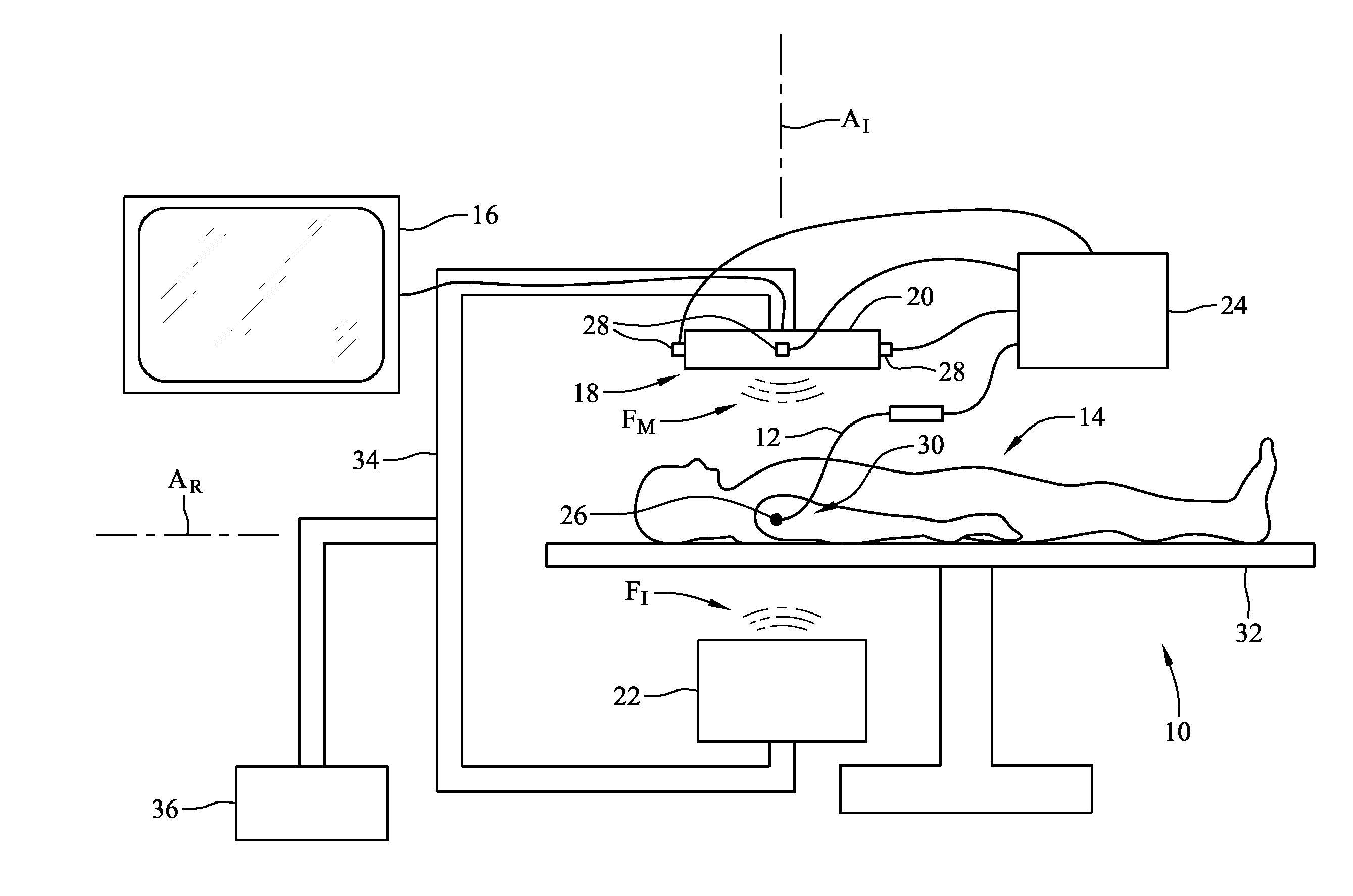
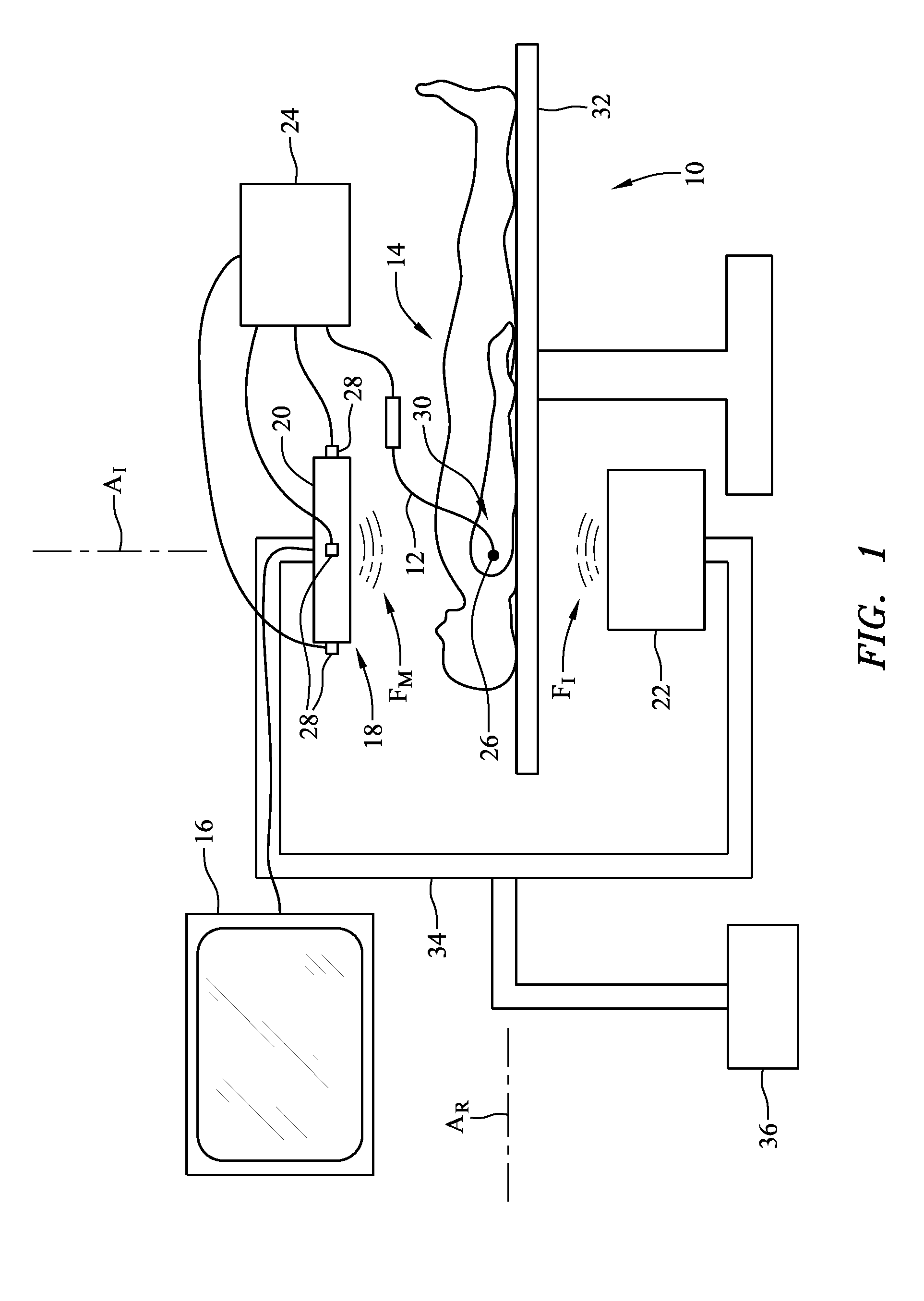
[0021]FIG. 1 is a schematic illustration of medical imaging system 10 for determining the position of catheter 12 relative to a 3D reconstructed model of an organ of patient 14, as well as for generating and displaying tracking-based enhancement information on display unit 16. System 10 includes moving imager 18, which includes intensifier 20 and emitter 22, and medical positioning system 24, which includes positioning sensor 26 and field generators 28. Electrophysiology map information and cardiac mechanical activation data pertaining to the model generated by medical imaging system 10 are displaye...


PUM


Abstract

A method for enhancing a three-dimensional (3D) reconstruction of an object comprises obtaining a signal indicative of a static 3D reconstruction of an object disposed in a tracking space, co-registering the 3D reconstruction to the 3D tracking space, collecting enhancement data from a tracked tool disposed in the 3D tracking space, and adding real-time features of the object to the static 3D reconstruction using the enhancement data. A system for enhancing data obtained by a medical system includes an electronic control unit configured to receive a first signal for a static 3D reconstruction of an organ, co-register the static 3D reconstruction to a 3D tracking space for a tracked tool, receive a second signal for enhancement data generated by the tracked tool operating within a region of interest of the organ, and add real-time features of the area of interest to the static 3D reconstruction using the enhancement data.

Description

CROSS-REFERENCE TO RELATED APPLICATIONS[0001]This application claims the benefit of U.S. provisional application No. 62 / 116,037, filed 13 Feb. 2015, which is hereby incorporated by reference as though fully set forth herein.BACKGROUND[0002]a. Field of the Disclosure[0003]The present disclosure relates generally to medical imaging and positioning systems that generate three-dimensional (3D) reconstructions of internal organs. In particular, the present disclosure relates to adding functional enhancements to 3D reconstructed models.[0004]b. Background Art[0005]Various methods exist for generating three-dimensional (3D) reconstructions of internal organs. For example, Computer Tomography (CT), X-ray, Positron Emission Tomography (PET) or Magnetic Resonance Imaging (MRI) may be used to generate a 3D modality that can be projected over fluoroscopy or some other two-dimensional (2D) image. Superimposing a real-time representation of an interventional medical device, such as a catheter or ...

Claims


Application Information

Patent Timeline
Patent Type & Authority Applications(United States)
IPC IPC(8): G06T7/00G06T11/00G06T7/20
CPCG06T7/0012A61B2034/2051G06T7/0081G06T7/20G06T11/003G06T2207/30048A61B6/12A61B6/4225A61B6/4441A61B6/481A61B6/503A61B6/5229A61B6/5264A61B5/042A61B5/063A61B34/20G06T7/004A61B34/10A61B90/37A61B90/39A61B2034/105A61B2090/376A61B2090/374A61B2576/023G06T7/70G06T7/11A61B2017/00243G16H30/40A61B5/283
Inventor KARIV, ITAY M.COHEN, AMITEICHLER, UZIKOR, KOBI
Owner ST JUDE MEDICAL INT HLDG SARL
Why Eureka
Try Eureka