Methods and devices for fallopian tube diagnostics

a fallopian tube and diagnostic technology, applied in the field of fallopian tube diagnostics, can solve the problems of disproportionately deadly ovarian cancer, low incidence of migration of ovarian cancer cells into the uterus, and inability to reliably diagnose ovarian malignancy by uterine sampling

Active Publication Date: 2016-09-29
BOSTON SCI SCIMED INC
View PDF6 Cites 30 Cited by
  • Summary
  • Abstract
  • Description
  • Claims
  • Application Information

AI Technical Summary

Problems solved by technology

Ovarian cancer is disproportionately deadly because this type of cancer lacks any clear early detection or screening test, meaning that most cases of ovarian cancer are not diagnosed until they have reached advanced stages.
Furthermore, biopsy of the ovary is not generally recommended by medical guidelines as there exists a risk of spreading the cancer further.
Cell samples obtained from the uterus may detect ovarian malignancy; however, the incidence of migration of ovarian cancer cells into the uterus is too low to render uterine sampling a reliable diagnostic test for ovarian malignancy.
However, the introduction of a diagnostic device into the Fallopian tube is problematic since the Fallopian tube is extremely fragile and prone to perforation during passage of most devices.

Method used


Image

  • Methods and devices for fallopian tube diagnostics
  • Methods and devices for fallopian tube diagnostics
  • Methods and devices for fallopian tube diagnostics

Examples


Embodiment Construction

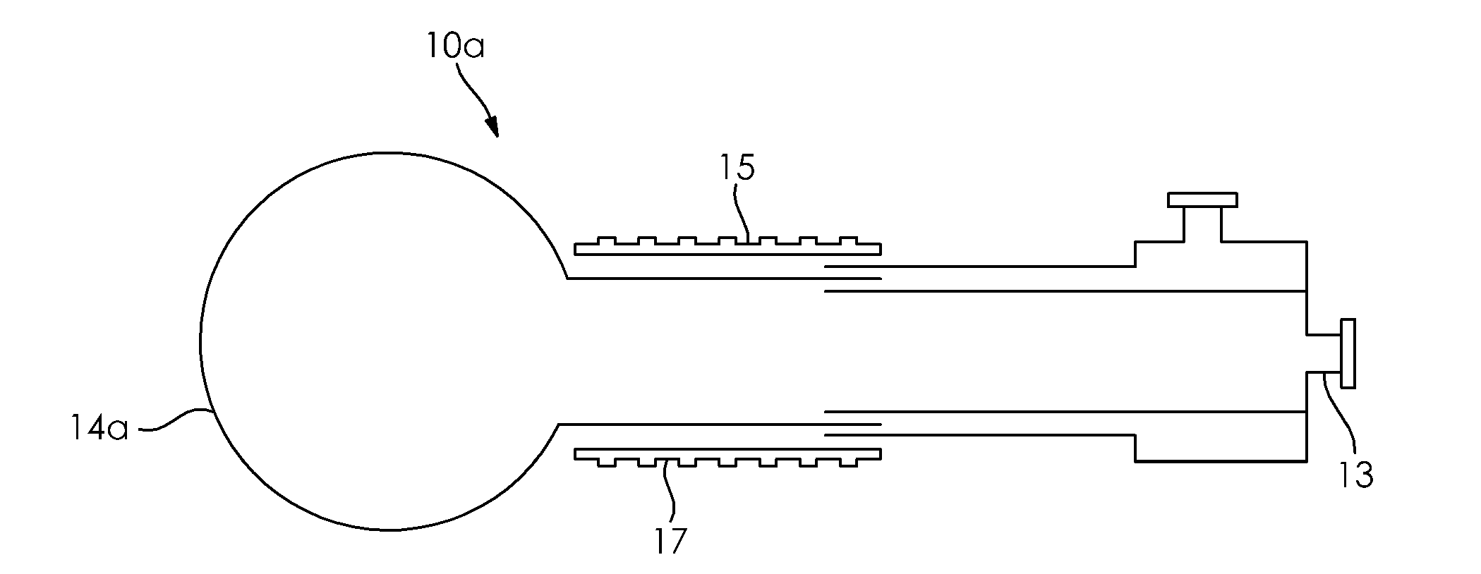
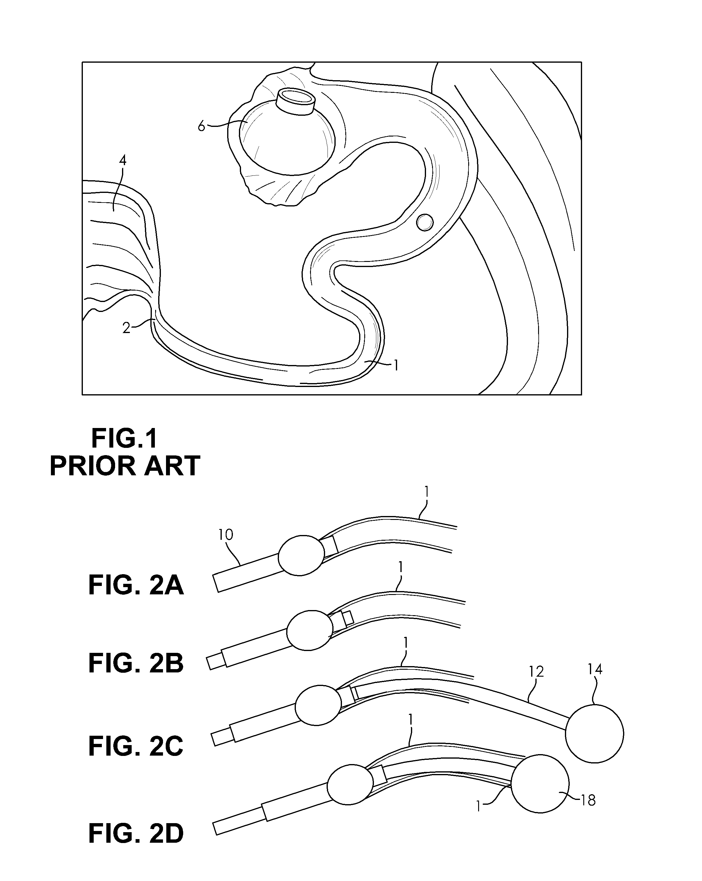
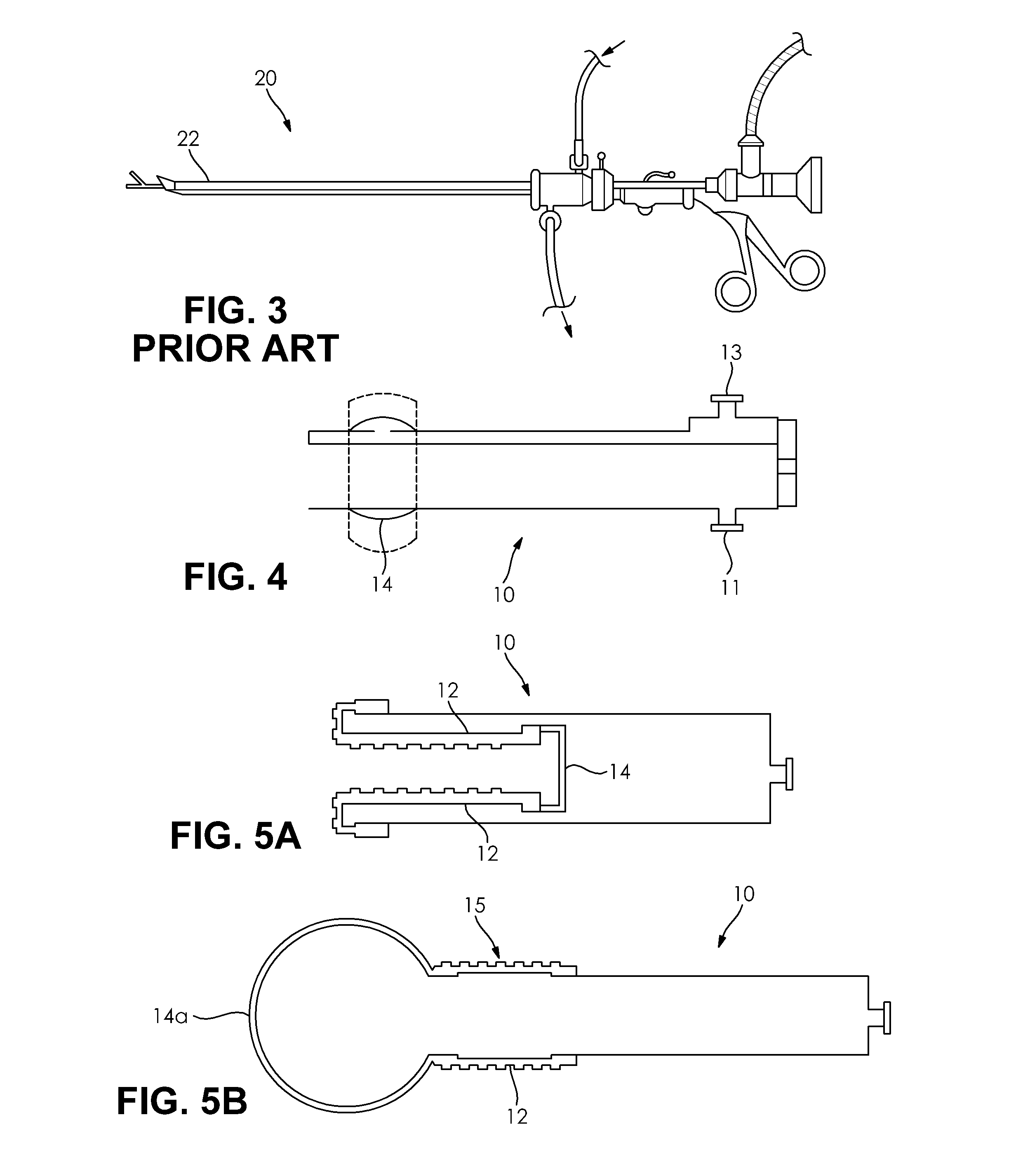
[0054]The present invention has utility in engaging the interior wall of the Fallopian tube and effectively removing cells therefrom for diagnostic purposes. A device and process is provided for collecting such cells in a minimally invasive procedure that in some embodiments occurs without cutaneous incision.

[0055]Where a range of values is provided, it is understood that each intervening value, to the tenth of the unit of the lower limit unless the context clearly dictates otherwise, between the upper and lower limits of that range is also specifically disclosed. Each smaller range between any stated value or intervening value in a stated range and any other stated or intervening value in that stated range is encompassed within the invention. The upper and lower limits of these smaller ranges may independently be included or excluded in the range, and each range where either, neither or both limits are included in the smaller ranges is also encompassed within the invention, subject...


PUM


Abstract

Methods and devices for performing minimally invasive procedures useful for Fallopian tube diagnostics are disclosed. In at least one embodiment, the proximal os of the Fallopian tube is accessed via an intrauterine approach; an introducer catheter is advanced to cannulate and form a fluid tight seal with the proximal os of the Fallopian tube; a second catheter inside the introducer catheter is provided to track the length of the Fallopian tube and out into the abdominal cavity; a balloon at the end of the second catheter is inflated and the second catheter is retracted until the balloon seals the distal os of the Fallopian tube; irrigation is performed substantially over the length of the Fallopian tube; and the irrigation fluid is recovered for cytology or cell analysis.

Description

RELATED APPLICATIONS[0001]This application is a continuation-in-part of U.S. patent application Ser. No. 14 / 764,710 filed Jul. 30, 2015; that in turn claims priority benefit of PCT / US14 / 14472 filed Feb. 3, 2014 that in turn claims priority benefit of U.S. Provisional Application Ser. No. 61 / 873,753 filed Sep. 4, 2013, and U.S. Provisional Application Ser. No. 61 / 759,783 filed Feb. 1, 2013; the contents of the aforementioned applications are hereby incorporated by reference in their entirety and incorporates the disclosure thereof in its entirety.FIELD OF THE INVENTION[0002]The present invention in general relates to Fallopian tube diagnostics and in particular to a catheter and diagnostic collection devices that accommodate the anatomical difficulties associated with navigation within the Fallopian tube.BACKGROUND OF THE INVENTION[0003]Ovarian cancer is a significant disease in women; 1 out of 72 women in the U.S. is diagnosed with ovarian cancer sometime during her lifetime. In 201...

Claims


Application Information

Patent Timeline
Patent Type & Authority Applications(United States)
IPC IPC(8): A61B10/02A61B1/303A61B90/00A61M25/10A61M25/01
CPCA61B10/0291A61M25/10A61M25/01A61M2025/1056A61B1/303A61B2090/3987A61M2025/105A61B90/39A61B2090/392A61B2090/3966A61B17/1204A61B17/12099A61B17/12136A61B2017/4233A61B2217/007
Inventor CHIN, ALBERTSARNA, SURBHISNOW, DAVID W.MAGANA, JESUS
Owner BOSTON SCI SCIMED INC
Why Eureka
Try Eureka