
Apparatus, systems and methods for determining tissue oxygenation

a tissue oxygenation and apparatus technology, applied in the field of apparatus, systems and medical devices, can solve the problems of inability to pain, need for reoperation, extreme sickness or death, and inability to reliably predict when a failure will occur

Inactive Publication Date: 2016-10-27
SURGISENSE CORP
View PDF8 Cites 201 Cited by
  • Summary
  • Abstract
  • Description
  • Claims
  • Application Information

AI Technical Summary

Benefits of technology

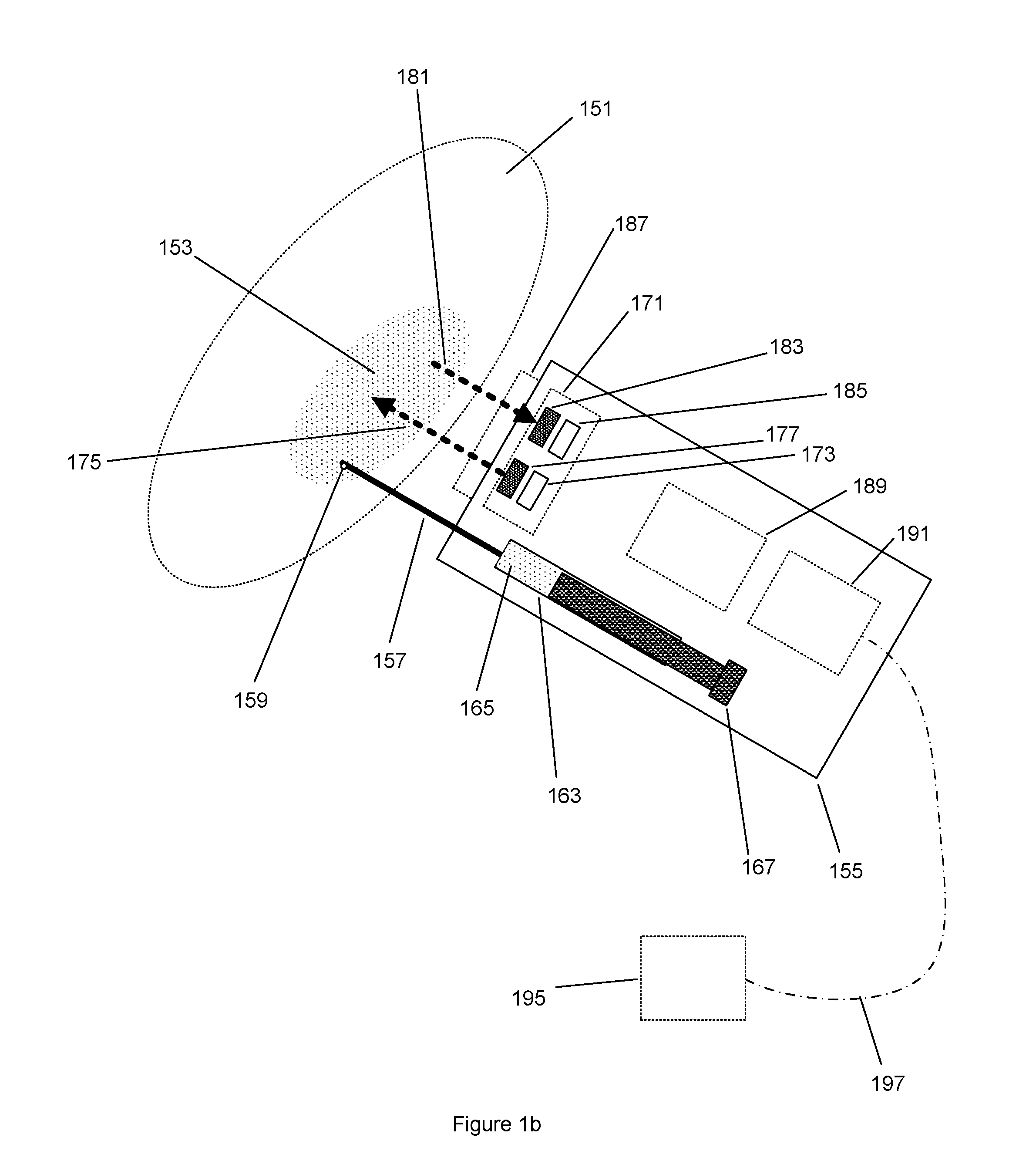
The patent describes a surgical instrument that can sense tissue oxygenation using a light re-emitting probe. The instrument includes an optical emitter to excite the probe, an optical detector to receive the re-emitted light, and a signal processor to resolve the tissue oxygenation based on the received light. The instrument can be used in a surgical stapler anvil or a monitoring device to map oxygenation of tissue. The technical effect of this invention is to provide a more accurate measure of tissue oxygenation during surgical procedures, which can improve the success of the surgery and reduce the risk of complications.

Problems solved by technology

Although many surgical procedures are successful, there is always a chance of failure.
Depending on the type of procedure these failures can result in pain, need for re-operation, extreme sickness, or death.
At present there is no reliable method of predicting when a failure will occur.
Most often the failure occurs after the surgical procedure has been completed.
The most difficult failures to predict and avoid are those that involve biological tissue.
Firstly, the properties that favor the continued function of biological tissue are very complex.
Secondly, these properties are necessarily disrupted by surgical manipulation.
However, traditional surgical instruments do not have the ability to obtain information from biological tissues.

Method used


Image

  • Apparatus, systems and methods for determining tissue oxygenation
  • Apparatus, systems and methods for determining tissue oxygenation
  • Apparatus, systems and methods for determining tissue oxygenation

Examples


Embodiment Construction

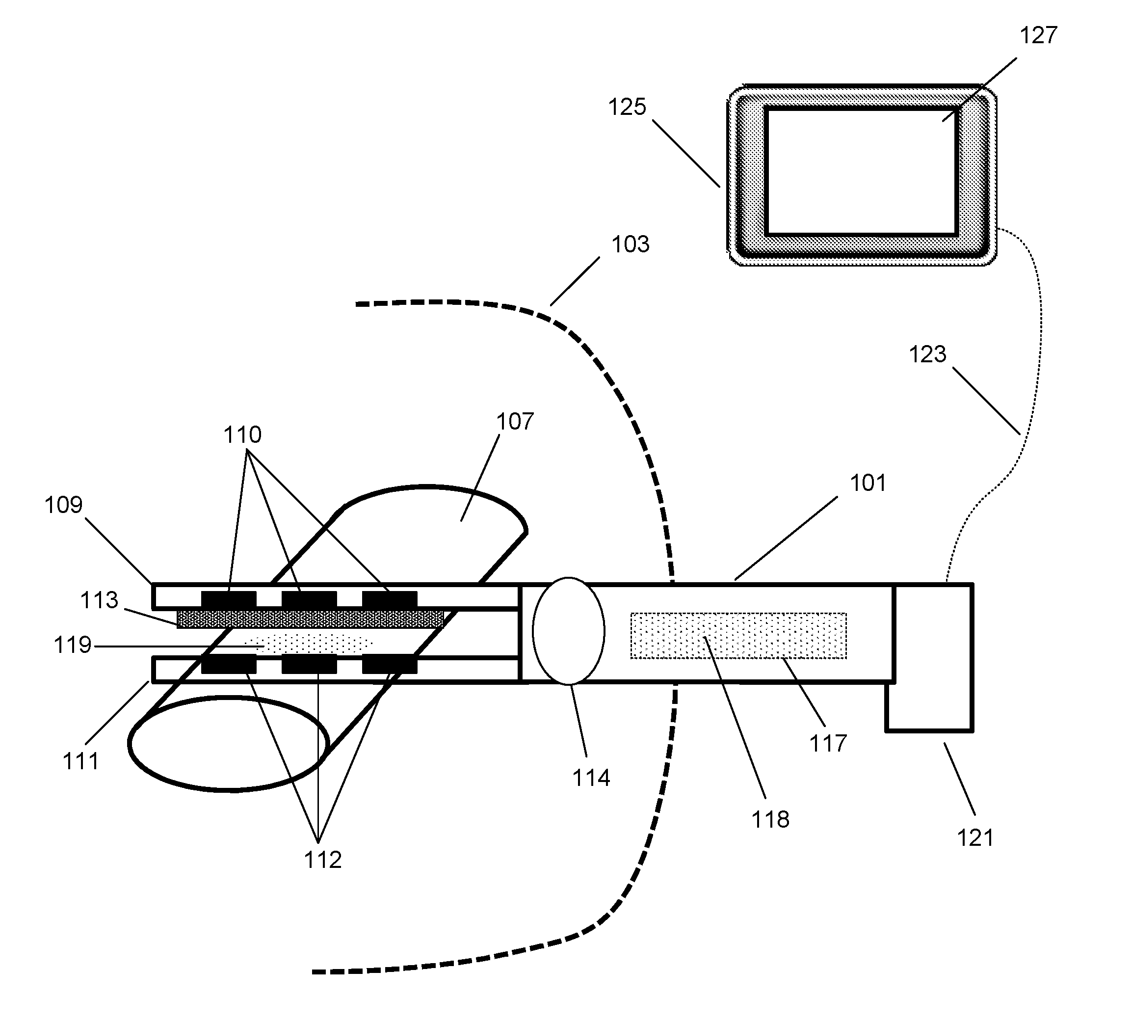
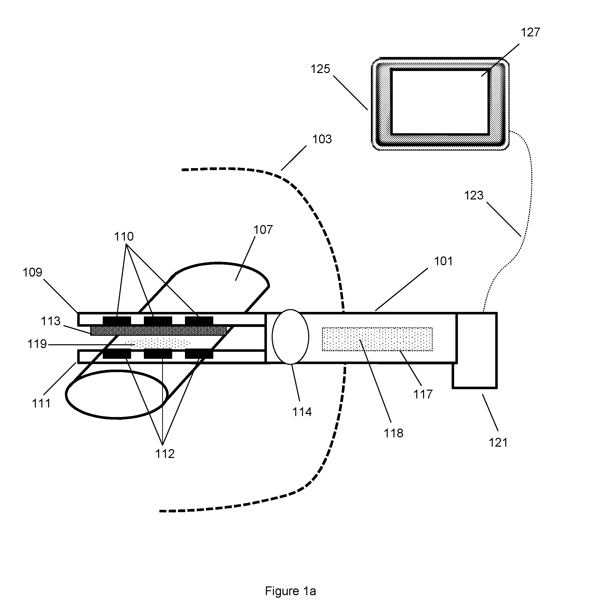
[0036]Each surgical procedure has the potential for failure. A common procedure in gastrointestinal surgery is a bowel resection—removing the affected portion of the bowel and then mechanically joining the ends of the remaining segments to re-establish bowel continuity. The mechanical connection of the free ends of bowel forms what is termed a surgical anastomosis. A surgical anastomosis is formed by either traditional techniques using suture material, or by contemporary techniques which may include utilizing surgical staplers or other surgical fastening device. A surgical stapler mechanically joins the bowel by firing a pattern of staples from a cartridge or housing through the two free ends of bowel against an anvil that ultimately forms a securing crimp on the opposing side. There are many embodiments of surgical staplers. Some staplers form linear staple patterns, while others form circular patterns. Some staplers incorporate functionality for cutting tissue. Many staplers have ...


PUM


Abstract

A surgical instrument may be configured to sense a light re-emitting probe to resolve tissue oxygenation, the surgical instrument including: an optical emitter configured to excite the light re-emitting probe within an absorption band of the light re-emitting probe; an optical detector configured to receive the re-emitted light from the probe; and a signal processor configured to resolve the tissue oxygenation based on the received light. The surgical instrument can be a surgical stapler anvil or a flexible substrate having a tissue interfacing surface. Further, a monitoring device may be configured to map oxygenation of a tissue containing a light re-emitting probe, the monitoring device including: an optical emitter configured to excite the light re-emitting probe; at least one optical detector configured to receive the re-emitted light from the probe; and a signal processor that is configured to resolve the tissue oxygenation at multiple points to generate an oxygen map.

Description

CROSS REFERENCE TO RELATED APPLICATIONS[0001]This application is a continuation of U.S. patent application Ser. No. 14 / 220,034 filed Mar. 19, 2014, which claims priority to U.S. Provisional Patent Application No. 61 / 803,451, filed Mar. 19, 2013, which is hereby incorporated herein by reference in its entirety.GOVERNMENT INTEREST[0002]This invention was made with Government Support under National Institutes of Health grant no. CA153571. The Government has certain rights in the invention.TECHNICAL FIELD[0003]The present invention relates to systems, methods and medical devices for use with molecular probes for resolving tissue oxygenation. More particularly, the inventions are directed to systems, methods and medical devices with sensors used to detect properties of biological tissue and a system for resolving the information gathered by the sensors.BACKGROUND[0004]A living organism is made up of cells. Cells are the smallest structures capable of maintaining life and reproducing. Cel...

Claims


Application Information

Patent Timeline
Patent Type & Authority Applications(United States)
IPC IPC(8): A61B5/1455A61B5/00A61B5/1459A61M5/00A61B17/068
CPCA61B5/14556A61B5/14552A61M5/007A61B17/068A61B2562/0238A61B5/4836A61B2017/00057A61B2090/065A61B2017/00084A61B5/1459A61B5/6847A61B2505/05A61B17/07207A61B17/1155A61B2017/07257A61B5/01A61B5/6868A61B5/0059
Inventor ZAND, JASON M.FISCHER, GREGORY S.
Owner SURGISENSE CORP
Learn More