Systems and methods for image/video recoloring, color standardization, and multimedia analytics
a technology of image/video recoloring and multimedia analytics, applied in image enhancement, image analysis, texturing/coloring, etc., can solve problems such as image misinterpretation, difficult color standardization, and adverse effects of classification performan
- Summary
- Abstract
- Description
- Claims
- Application Information
AI Technical Summary
Benefits of technology
Problems solved by technology
Method used
Image
Examples
example iii
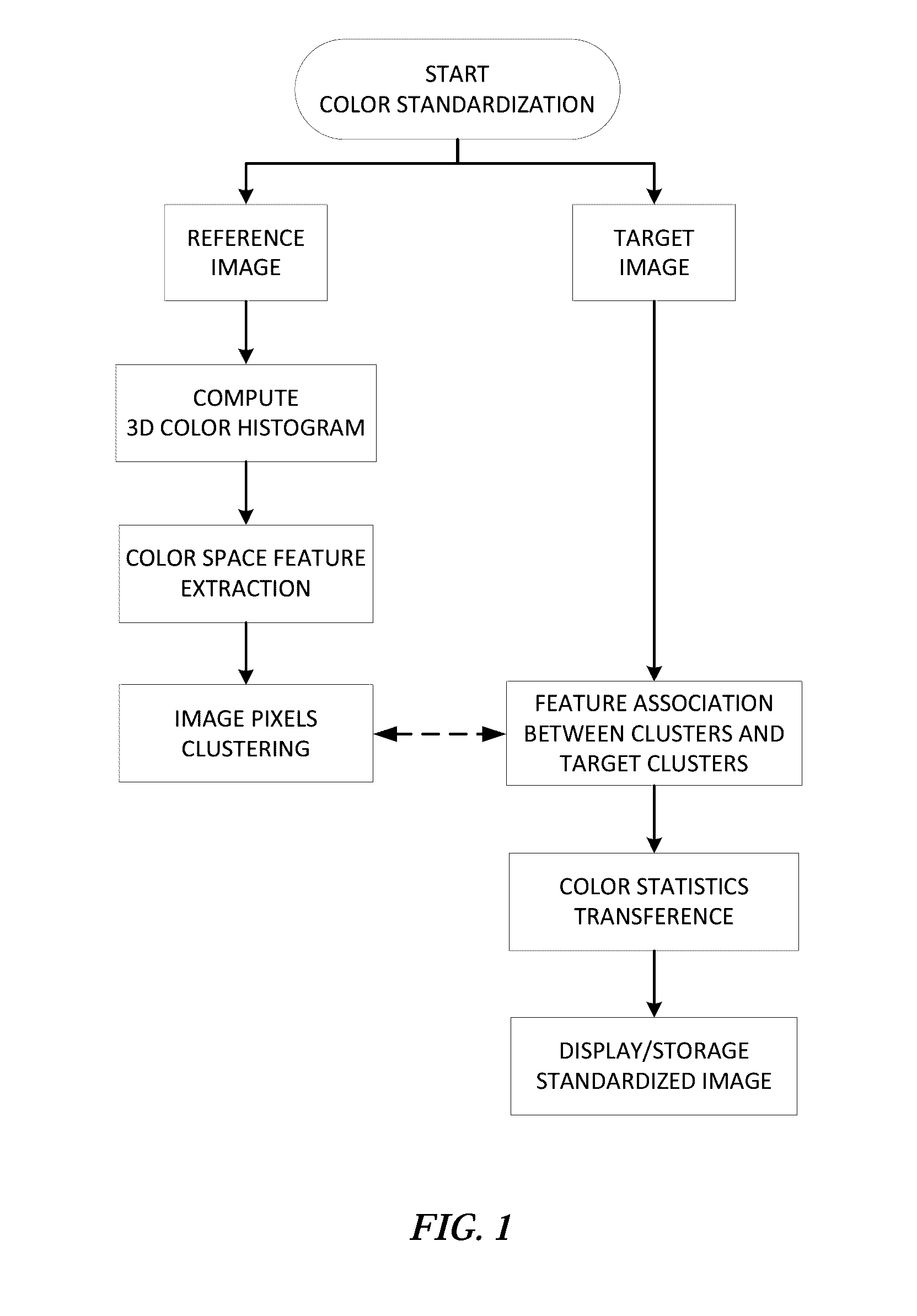
[0064]1. Compute the 3D color histogram for both the reference and input images.
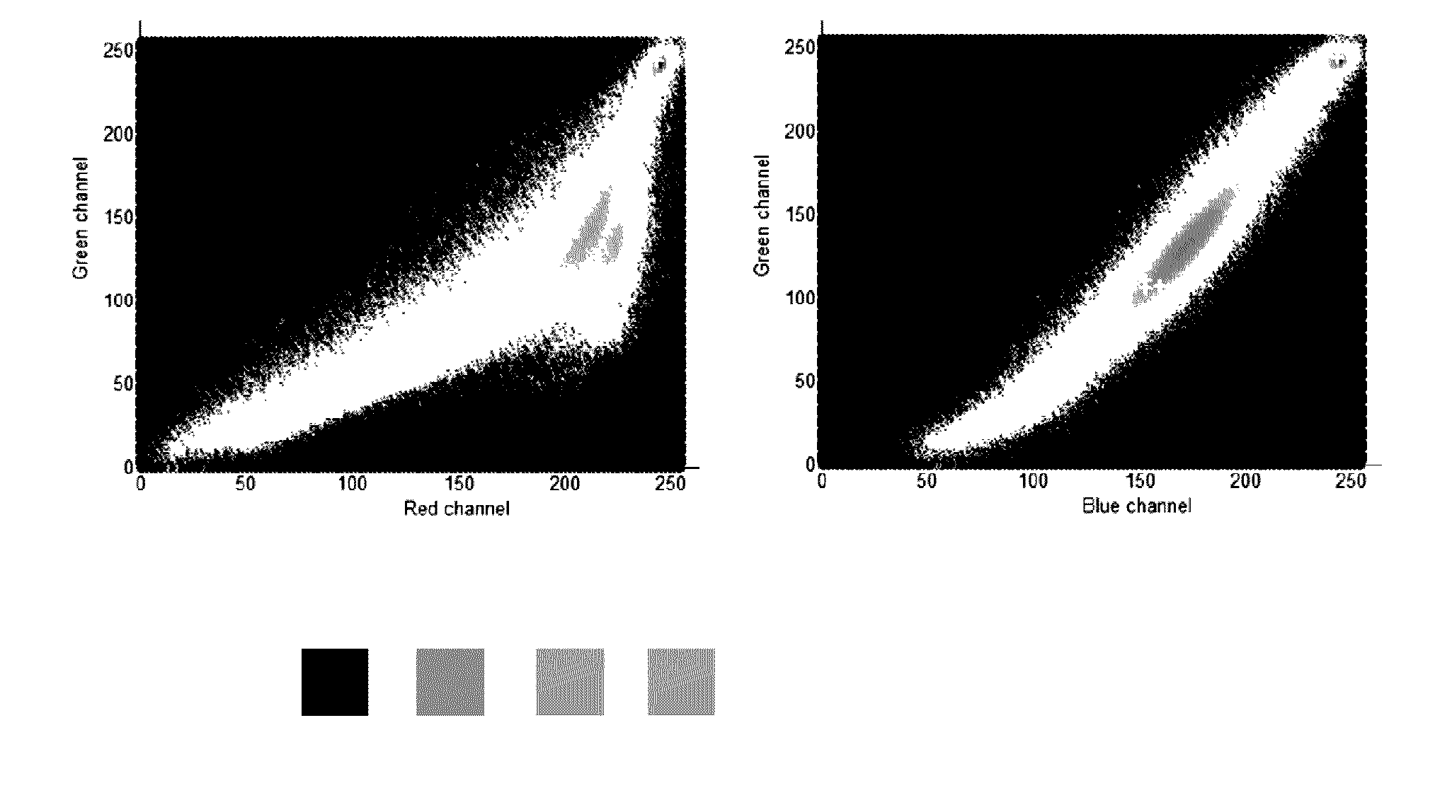
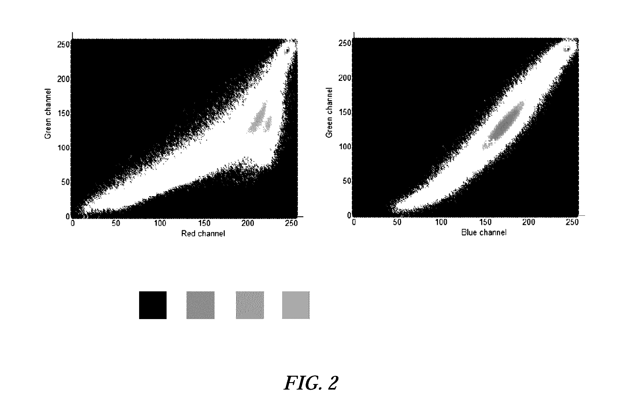
[0065]2. Find the n more relevant colors in the reference and input images using scale space maxima detection in the 3D color histogram.
[0066]3. Find the average and entropy of the more relevant colors.
[0067]4. Compare the resulting colorfulness measure between reference and input image.
In addition to the aforementioned colorfulness measure, additional examples of measures that can be used for colorfulness quantitative analysis may be found in these references: A Othman and K. Martinez, Colour appearance descriptors for image browsing and retrieval, PROC. SPIE ELECTRONIC IMAGING 2008, 2008; G. Wyszecki and W. Stiles, Color science: Concepts and methods, quantitative data and formulae, New York, NY: John Wiley & Sons, 1982; C. Gao, K. Panetta and S. Agaian, Color image attribute and quality measurements, PROC. SPIE SENSING TECHNOLOGY+APPLICATIONS, 2014.;K. Panetta, C. Gao and S. Agaian, No reference Color...
example i
Color Standardization of Tissue Microarray Cores of Prostate Tissue
[0069]A batch of 360 H&E-stained tissue microarray cores of prostate tissue were standardized using the disclosed method. Local standardization of image pixels associated with broad prostate tissue structures (e.g., lumen, stroma, and epithelium) is carried out having as reference image the image shown in FIG. 3. The following steps were executed: (1) unsupervised color space feature extraction using scale space maxima detection for defining reference colors from a well-stained histopathology slide, (2) feature association between the resulting reference clusters and target clusters, and (3) weighted linear mapping of statistical moments (mean and standard deviation) from the reference image to target images. The standardized images are tested for assessing color consistency using normalized median intensity (NMI) (the lower the standard deviation of the NMI the better the color constancy) from segmented regions, and...
example ii
Face Color Transference
[0070]The example in FIG. 6 shows the results obtained using the fuzzy color transference in skin color lightening. The reference skin color centroid is detected by finding the most frequent color (3D histogram global maximum) within the skin area previously selected by the user. In the same way the centroid color are found for the target image. Since the only cluster in this example is the face, the mean and standard deviation of the face pixels are computer for each channel and for both reference and target image. Then, color transference is performed from the reference to the target image using the following equation:
Iinorm,ch(x,y)=Iich(x,y)+u(x,y)j((α-μIr,jch)+σIr,jchσIi,jch(Iich(x,y)-(α-μIi,jch))-Iich(x,y))
In this example, the color model using for color processing is lab color space. Once all transformation a performed in a per-pixel basis, image pixels in the target image are converted to RGB for visualization or storage.
Additional Embodiments
[0071]The ...
PUM
 Abstract
Description
Claims
Application Information
 - R&D
- Intellectual Property
- Life Sciences
- Materials
- Tech Scout
- Unparalleled Data Quality
- Higher Quality Content
- 60% Fewer Hallucinations
Browse by: Latest US Patents, China's latest patents, Technical Efficacy Thesaurus, Application Domain, Technology Topic, Popular Technical Reports.
© 2025 PatSnap. All rights reserved.Legal|Privacy policy|Modern Slavery Act Transparency Statement|Sitemap|About US| Contact US: help@patsnap.com


