Method for generating a 3D reference computer model of at least one anatomical structure

Inactive Publication Date: 2016-11-17
AO TECH AG
View PDF2 Cites 24 Cited by
  • Summary
  • Abstract
  • Description
  • Claims
  • Application Information

AI Technical Summary

Benefits of technology

[0060]A preferred use of the method is for the quality assurance of surgical treatments. A further component and advantage of this method is that all data that is generated over the entire course of therapy can be integrated into

Problems solved by technology

A disadvantage of this known method can be that each a CT of the

Method used


Image

  • Method for generating a 3D reference computer model of at least one anatomical structure
  • Method for generating a 3D reference computer model of at least one anatomical structure
  • Method for generating a 3D reference computer model of at least one anatomical structure

Examples


example 1

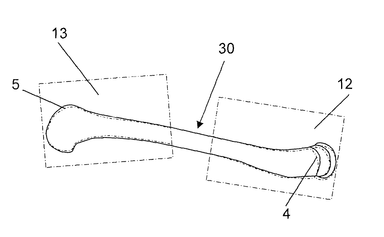
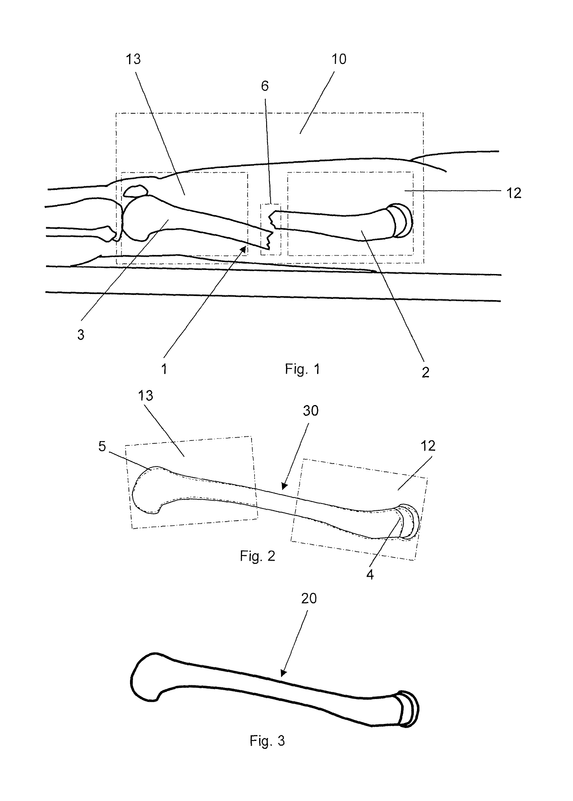
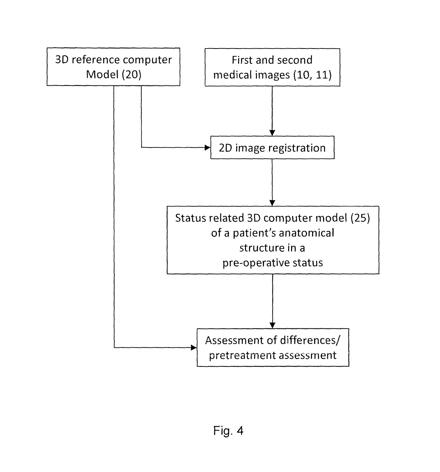
[0082]Hereinafter, the method for generating a 3D reference computer model 20 according to the invention, the method for generating a status related 3D computer model 25 according to the invention and the method for generating a graphical 3D computer model 21 are described at an example of a surgical treatment of bone fractures and a correction of osseous deformities.

[0083]First, preoperative first and second medical images 10, 11 of the anatomical structures of a patient to be treated are acquired by means of a computer-aided medical imaging procedure. The method includes obtaining adequate image information of the operation area prior to surgery. The method provides acquiring a preoperative first medical image data set of an anatomical structure of a patient to be treated, preferably using a CT, for example the region with a bone fracture or osseous deformity. Alternatively, or in addition other 3D layer imaging techniques such as cone beam computed tomography can (called digital ...

example 2

[0120]The method for generating a 3D reference computer model 20 according to the invention, the method for generating a status related 3D computer model 25 according to the invention and the method for generating a graphical 3D computer model 21 are described below at another example for applications in the dental implantology. The course of therapy in the case of implantation of one or more dental implants can be monitored over the course of the therapy as follows: preoperatively at least a first and second medical image 10, 11 of the operation area and the neighbouring region, e.g. around the adjacent teeth and / or of the alveolar ridge are acquired, i.e. a preoperative medical 3D image data set is obtained 10 and a 3D reference computer model 25 and / or a pre-operative status related 3D computer model 25 and / or a sub model thereof is generated. Preferably, the 3D imaging is performed using an optical 3D scanning procedure, e.g. laser scanning. This 3D imaging can be effected solel...


PUM


Abstract

A method for generating a 3D reference computer model of at least one anatomical structure for comparison with a selectable pre-, intra- or postoperative set of medical images of at least one anatomical structure, the method including: acquiring at least a first and a second medical image of at least one anatomical structure in a preoperative status from different perspectives using a computer assisted medical imaging device, wherein the first and second medical images are represented by a respective first and second set of digital 2D image data; and generating a 3D reference computer model of an anatomical structure by selecting and extracting a 3D atlas model of an anatomical structure to be treated from a generic anatomical atlas provided in the form of a digital data source, and registering at least a section of each of the first and second medical images to the selected 3D atlas model.

Description

BACKGROUND OF THE INVENTION[0001]1. Field of the Invention[0002]The invention relates to a method for generating a 3D reference computer model of at least one anatomical structure according to the preamble of claim 1, to a method for generating a status related 3D computer model of a patient's anatomical structure in a pre-, intra- or post-operative status according to the preamble of claim 10 or 11 and to a method for monitoring a surgical treatment according to the preamble of claim 26.[0003]During surgical treatments of fractures and the correction of osseous deformities bone fragments are anatomically repositioned and stably fixed at a correct position by using suitable fixation techniques. Problems may arise by an unrecognized malposition of bone fragments and implants during surgery, or through their secondary dislocation in the postoperative course. A faulty osteosynthesis due to anatomically incorrect repositioning of bone fragments, improper surgical technique, unsuitable s...

Claims


Application Information

Patent Timeline
IPC IPC(8): A61B34/10G06T19/20G06T7/00G06T17/00
CPCG06T2207/10004A61B2034/101A61B34/10G06T17/00G06T2207/20104G06T7/0032G06T2210/41G06T2207/30008G06T2207/20101G06T19/20A61B2034/104A61B2034/105A61B2090/367A61B2090/376A61B2090/3762G06T2207/10116G06T2207/20128G06T7/344
Inventor NOTZLI, CHRISTOPHKAMER, LUKAS
Owner AO TECH AG
Why Eureka
Try Eureka