
Group sparsity model for image unmixing

a group sparsity model and image unmixing technology, applied in image enhancement, image analysis, instruments, etc., can solve the problem of spatial continuity loss in unmixed images, the limitation of the maximum number of stains that can be solved, and the limitation of the number of large stains. problem, to achieve the effect of reducing the model to sparse unmixing

Active Publication Date: 2016-12-08
VENTANA MEDICAL SYST INC
View PDF7 Cites 8 Cited by
  • Summary
  • Abstract
  • Description
  • Claims
  • Application Information

AI Technical Summary

Benefits of technology

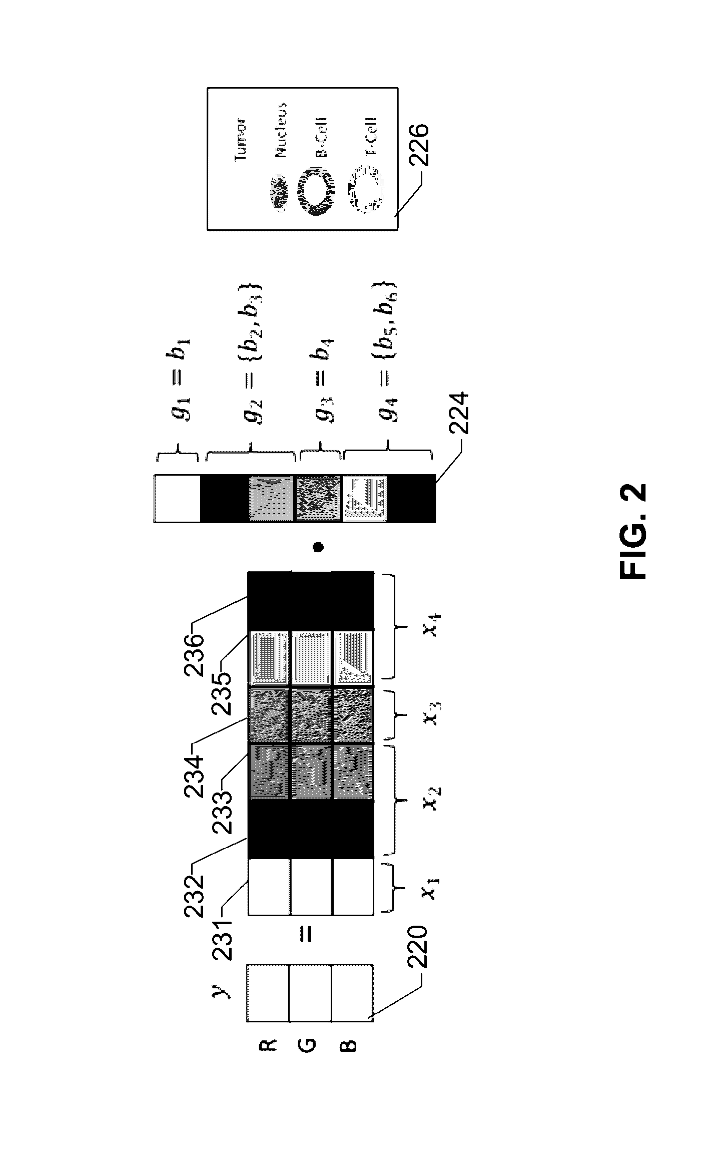
The present invention provides a system and method for unmixing multiple stain contributions in a tissue image, which is useful for analyzing the composition of cells or tissues. The approach involves using a group sparsity framework and a co-localization information model to isolate the contributions of different stains and solving the model using a group lasso algorithm to yield a least squares solution for each group. This method, which can be implemented using a computer-implemented method or a system, provides a more accurate and efficient way to unmix images than previous techniques and can help to improve the analysis of tissue samples.

Problems solved by technology

This technique is most widely used in the current digital pathology domain, however, the maximum number of stains that can be solved is limited to three as the linear system is deficient for not enough equations (X being a 3×M matrix).
This method also solves for y=Xb and has the same limitation in dealing with large stain numbers.
Due to the independent assignment of each pixel into different systems, the spatial continuity is lost in the unmixed images and artifacts such as holes are observed.
Sparse models for high dimensional multi-spectral image unmixing adopt the L0 norm to regularize the combination weights b of the reference colors hence leading to a solution that only a small number of reference colors are contributed to the stain color mixture, but these are also designed for multi-spectral images and do not use any prior biological information about the biomarkers, which may lead to undesired solutions for real data.
Moreover, these methods cannot be applied to RGB images due to the image acquisition system, i.e. multi-spectral imaging instead of a CCD color camera to capture the image using a set of spectral narrow-band filters.
The linear system constructed from it is always an over-determined system with X being a K×M(K>>M) matrix that leads to a unique solution, however, the scanning process in the multi-spectral imaging system is very time consuming and only a single field of view manually selected by a technician can be scanned instead of the whole slide, thereby limiting the usage of such methods.
Therefore, there exists no numerical solution for unmixing an image having more unknown variable than the number of equations in the least squares system.
Due to the limitations of a CCD color camera, an acquired RGB or brightfield image only contains three channels, the unmixing of which into more than three colors is a challenging task.

Method used


Image

  • Group sparsity model for image unmixing
  • Group sparsity model for image unmixing
  • Group sparsity model for image unmixing

Examples


Embodiment Construction

[0025]Before elucidating the embodiments shown in the Figures, various embodiments of the present disclosure will first be described in general terms.

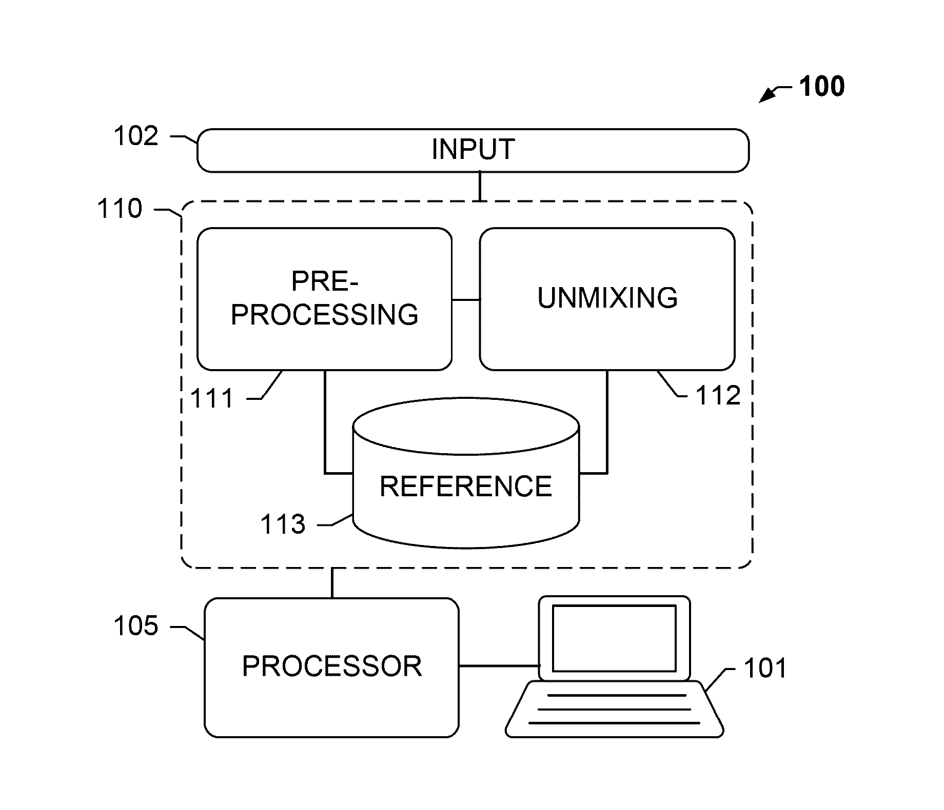
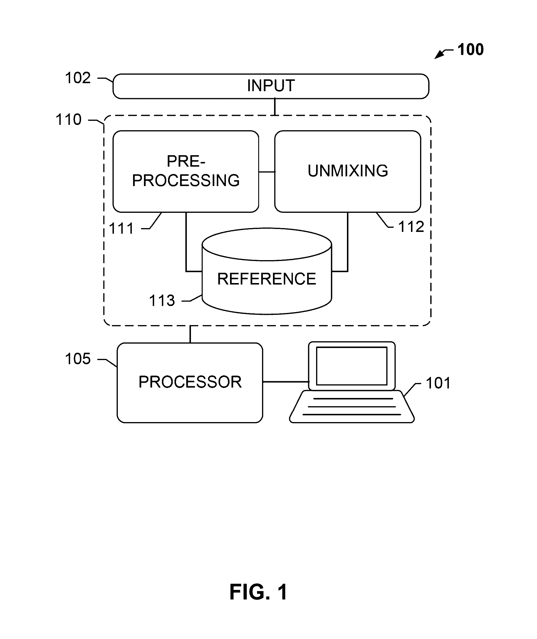
[0026]The present disclosure relates, inter alia, to an analysis system, e.g. to a tissue analysis system. The system may be suitable for analyzing biological specimen, for example, tissue provided on a slide.

[0027]The term “image data” as understood herein encompasses raw image data acquired from the biological tissue sample, such as by means of an optical sensor or sensor array, or pre-processed image data. In particular, the image data may comprise a pixel matrix.

[0028]The analysis system may comprise a color data storage module that stores color data, e.g. color data indicative of a color of the stains. Such color data is also referred to as “reference data”. The color data may be descriptive of a single frequency or a characteristic spectral profile of the stain. The color data storage module may store color data for each of a plu...


PUM


Abstract

Systems and methods described herein relate, among other things, to unmixing more than three stains, while preserving the biological constraints of the biomarkers. Unlimited numbers of markers may be unmixed from a limited-channel image, such as an RGB image, without adding any mathematical complicity to the model. Known co-localization information of different biomarkers within the same tissue section enables defining fixed upper bounds for the number of stains at one pixel. A group sparsity model may be leveraged to explicitly model the fractions of stain contributions from the co-localized biomarkers into one group to yield a least squares solution within the group. A sparse solution may be obtained among the groups to ensure that only a small number of groups with a total number of stains being less than the upper bound are activated.

Description

CROSS-REFERENCE TO RELATED APPLICATIONS[0001]This patent application is a continuation of International Patent Application No. PCT / EP2015 / 053745 filed Feb. 23, 2015, which claims priority to and the benefit of U.S. Provisional Patent Application Ser. No. 61 / 943,265, filed Feb. 21, 2014. Each patent application is incorporated herein by reference as if set forth in its entirety.BACKGROUND OF THE SUBJECT DISCLOSURE[0002]Field of the Subject Disclosure[0003]The present subject disclosure relates to digital pathology. More particularly, the present subject disclosure relates to color unmixing methods and systems for a multiplex IHC image that can accommodate any number of stain colors.[0004]Background of the Subject Disclosure[0005]Multiplex immunohistochemistry (IHC) staining is an emerging technique for the detection of multiple biomarkers within a single tissue section and has become more popular due to its significant efficiencies and the rich diagnostic information it has. A multip...

Claims


Application Information

Patent Timeline
Patent Type & Authority Applications(United States)
IPC IPC(8): G06T7/00G06K9/00G06T7/40
CPCG06T7/0012G06T7/408G06K9/00147G06T2207/30204G06T2207/10056G06T2207/30024G06T2207/10024G06V20/698G06T7/90
Inventor CHUKKA, SRINIVASCHEN, TING
Owner VENTANA MEDICAL SYST INC
Learn More