
Model-based segmentation of an anatomical structure

a model-based, anatomical technology, applied in the field of model-based segmentation of anatomical structures, can solve problems such as insufficient suitability, and achieve the effects of improving spatial resolution of deformable models, improving imaging, and improving depth precision

Active Publication Date: 2016-12-29
KONINKLJIJKE PHILIPS NV
View PDF13 Cites 4 Cited by
  • Summary
  • Abstract
  • Description
  • Claims
  • Application Information

AI Technical Summary

Benefits of technology

The invention is a method for creating a flexible model that can better segment the walls of an anatomical structure in a medical image. This is done by creating a layer of mesh that matches the topologies of two surface layers, which helps to accurately match the composition of the walls. The result is a more detailed and accurate model for medical purposes.

Problems solved by technology

A problem of the known deformable models is that they are insufficiently suitable for providing a detailed segmentation of a wall of such anatomical structures.

Method used


Image

  • Model-based segmentation of an anatomical structure
  • Model-based segmentation of an anatomical structure
  • Model-based segmentation of an anatomical structure

Examples


Embodiment Construction

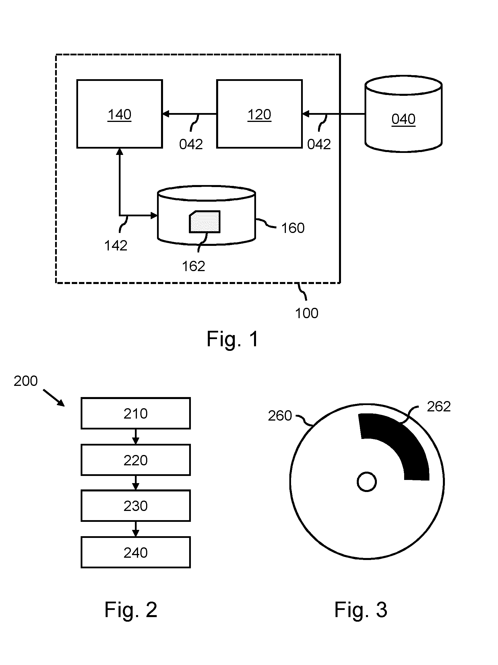
[0059]FIG. 1 shows a system for generating a deformable model for segmenting an anatomical structure in a medical image, as well as a system for applying the deformable model to the anatomical structure in the medical image. By way of example, FIG. 1 shows a single system 100 providing both functionalities. It will be appreciated, however, that in practice both functionalities may be separated, i.e., performed by different systems.

[0060]Referring firstly to the generating of the deformable model, the system 100 may comprise a processing subsystem 140 configured for:

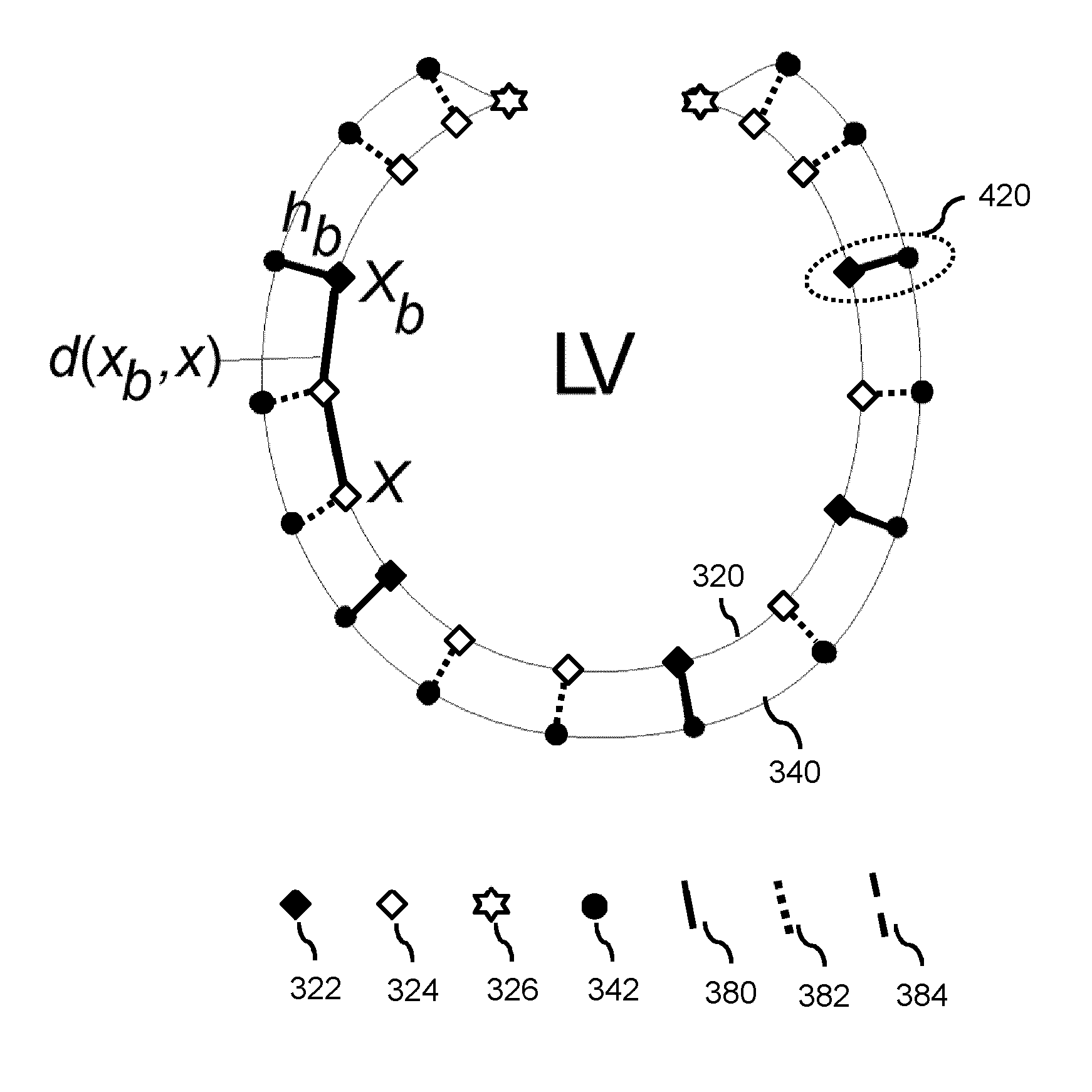
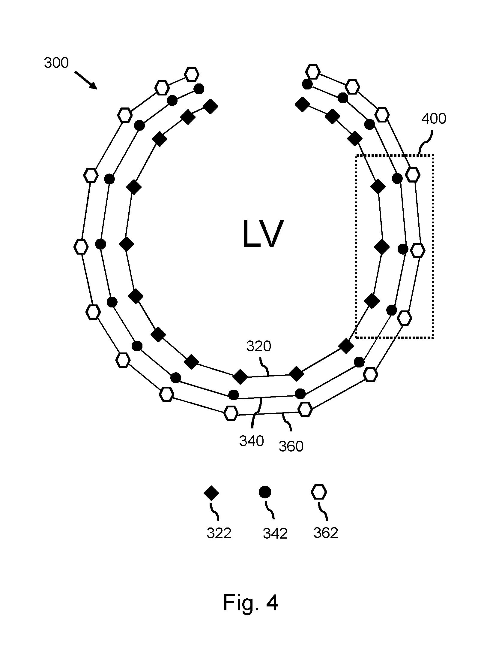
i) providing a first surface mesh for being applied to the first surface layer of the wall during a model-based segmentation,

ii) providing a second surface mesh for being applied to the second surface layer of the wall during the model-based segmentation, and

iii) generating an intermediate layer mesh for being applied in-between the first surface layer and the second surface layer during the model-based segmentation, said...


PUM


Abstract

A method is provided for generating a deformable model (300) for segmenting an anatomical structure in a medical image. The anatomical structure comprises a wall. The deformable model (300) is generated such that it comprises, in addition to two surface meshes (320, 360), an intermediate layer mesh (340) for being applied in-between a first surface layer of the wall and a second surface layer of the wall. In generating the intermediate layer mesh (340), the mesh topology of at least part (400) of the intermediate layer mesh is matched to the mesh topology of one of the surface meshes (320, 360), thereby establishing matching mesh topologies. The deformable model (300), as generated, better matches the composition of such walls, thereby providing a more accurate segmentation.

Description

FIELD OF THE INVENTION[0001]The invention relates to a method for generating a deformable model for segmenting an anatomical structure in a medical image. The invention further relates to model data being obtainable by the method. The invention further relates to a system for applying the deformable model to the anatomical structure in the medical image.BACKGROUND OF THE INVENTION[0002]Robust automatic segmentation of various anatomical structures in a medical image is a key enabler in improving clinical workflows. Here, the term segmentation refers to the identification of the anatomical structure in the medical image by, e.g., delineation of the boundaries of the anatomical structure, or by labeling of the voxels enclosed by the boundaries. Once such segmentation has been performed, it is possible to extract clinical parameters such as, in case of a cardiac structure, ventricular mass, ejection fraction and wall thickness. Consequently, automatic segmentation can significantly red...

Claims


Application Information

Patent Timeline
IPC IPC(8): G06T7/00A61B5/05
CPCG06T7/0089G06T7/0083G06T2207/10028G06T2207/10072G06T2207/30048G06T7/12G06T7/149A61B5/05A61B6/03G06T7/10
Inventor GROTH, ALEXANDRANICKISCH, HANNESWEBER, FRANK MICHAELWEESE, JUERGENBARSCHDORF, HANS
Owner KONINKLJIJKE PHILIPS NV
Learn More