Socs mimetics for the treatment of diseases
- Summary
- Abstract
- Description
- Claims
- Application Information
AI Technical Summary
Benefits of technology
Problems solved by technology
Method used
Image
Examples
example 1
SOCS1+ / − Mice Exhibit CD4 T Cell Accumulation and Enhanced CD4+ T Cell Activation in the Periphery
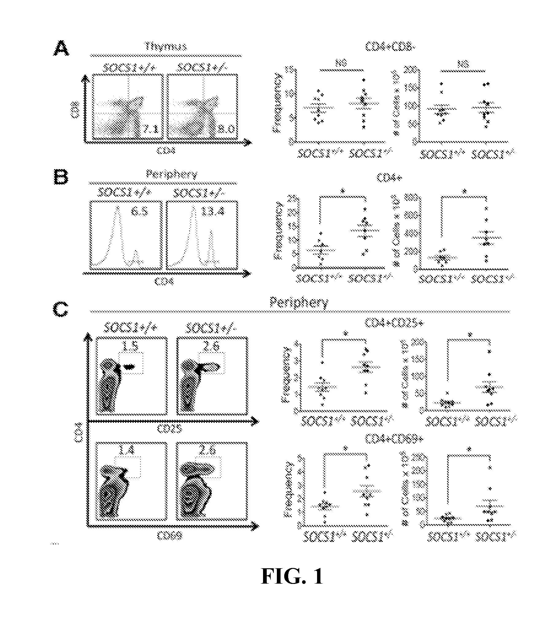
[0145]In this study we selected SOCS1+ / − mice between 2 and 5 months of age, which are free of overt lupus pathology (FIG. 7 and Fujimoto et al. 2004), to assess how SOCS1 deficiency precipitates CD4+ T lymphocyte mediated autoimmunity. We first assessed whether distinctions were present within the thymic and peripheral pools of CD4+ T lymphocytes in SOCS1+ / − mice. Although the frequency and absolute numbers of CD4+CD8− thymocytes within SOCS1+ / − and WT littermate controls were indistinct (FIG. 1A), CD4+ T lymphocytes in the periphery of SOCS1+ / − mice were two fold higher in frequency, and four-fold higher in absolute cell number, when compared to WT littermate controls (FIG. 1B). As T lymphocyte activation may also contribute to lupus progression (Stekman et al. 1991; Desai-Mehta et al. 1996; Mohan et al. 1999; Vratsanos et al. 2001; Bouzahzah et al. 2003), we next assessed differences...
example 2
CD28 Co-Stimulation of SOCS1+ / − CD4+ T Lymphocytes is Dispensable for Activation and Clonal Expansion
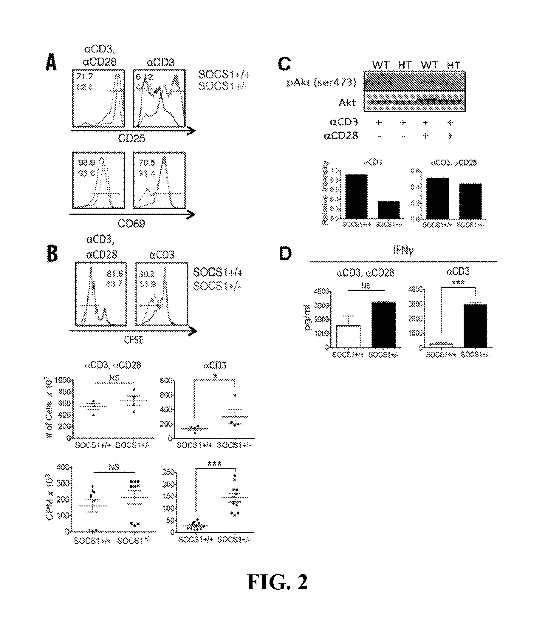
[0146]Having observed enhanced CD4+ T cell activation within the periphery of SOCS1+ / − mice, we next conducted in vitro assays to characterize this observation mechanistically. Given that conventional T lymphocytes (Tcon) require signaling through both TCR and CD28 for activation (reviewed in Tivol et al. 1996), we next stimulated MACs purified, CD4+CD25− T lymphocytes with antibodies specific to CD3 and CD28 (αCD3 / αCD28), in vitro. After 72 hours of incubation, αCD3 / αCD28 activated CD4+ T lymphocyte populations were greater than 70% CD25+, independent of SOCS1 expression (FIG. 2A). In similar fashion, αCD3 / αCD28 treatment mediated comparable CD69 up-regulation in both SOCS1+ / − and littermate control T lymphocytes (FIG. 2A). Notably, however, whereas αCD3 treatment mediated limited CD25 high expression in SOCS1 sufficient CD4+ T cells, 45% of SOCS1+ / − counterparts had high levels of ...
example 3
SOCS1+ / − CD4+ T Cells Exhibit Reduced P-Akt Requirement for Activation
[0148]As SOCS1+ / −CD4+ T lymphocytes exhibited significant activation and proliferation in the absence of CD28 stimulation, we next examined Akt activation, which is involved in signaling downstream of CD28 (Parry et al. 1997). Strikingly, although we observed significantly more proliferation and activation of SOCS1+ / − CD4+ T lymphocytes under αCD3 treatment alone, Akt phosphorylation subsequent to αCD3 treatment was significantly reduced in SOCS1+ / − mice compared to littermate controls (FIG. 2C). The differences in p-Akt were observed in spite of comparable amounts of total Akt under αCD3 treatment (FIG. 2C). Consistent with the activation and proliferation results noted previously (FIG. 2A, FIG. 2B), p-Akt levels were indistinct between SOCS1+ / − and WT cells incubated with αCD3 / αCD28 (FIG. 2C). Together, these results suggest a limited requirement for CD28 co-stimulatory signals in the CD4+ T lymphocyte phenotype...
PUM
| Property | Measurement | Unit |
|---|---|---|
| Level | aaaaa | aaaaa |
| Disorder | aaaaa | aaaaa |
| Lipophilicity | aaaaa | aaaaa |
Abstract
Description
Claims
Application Information
 - R&D
- Intellectual Property
- Life Sciences
- Materials
- Tech Scout
- Unparalleled Data Quality
- Higher Quality Content
- 60% Fewer Hallucinations
Browse by: Latest US Patents, China's latest patents, Technical Efficacy Thesaurus, Application Domain, Technology Topic, Popular Technical Reports.
© 2025 PatSnap. All rights reserved.Legal|Privacy policy|Modern Slavery Act Transparency Statement|Sitemap|About US| Contact US: help@patsnap.com


