Methods and Devices for Imaging Large Intact Tissue Samples

a tissue sample and large-scale technology, applied in the field of methods and devices for imaging large-scale intact tissue samples, can solve the problems of slow imaging speed of confocal microscopes and damage to the signal-emitting capabilities of samples

Active Publication Date: 2017-03-09
THE BOARD OF TRUSTEES OF THE LELAND STANFORD JUNIOR UNIV
View PDF4 Cites 14 Cited by
  • Summary
  • Abstract
  • Description
  • Claims
  • Application Information

AI Technical Summary

Benefits of technology

[0003]Methods and devices for conducting high-speed, high-resolution imaging of large intact tissue samples are provided. Aspects of the methods include placing a sample in an optically homogenous sample manipulation component, performing a calibration procedure to align a light sheet and a detection focal

Problems solved by technology

A major challenge to the imaging of large intact tissue samples is to conduct the imaging process in a practically feasible period of time.
Confocal microscopes

Method used


Image

  • Methods and Devices for Imaging Large Intact Tissue Samples
  • Methods and Devices for Imaging Large Intact Tissue Samples
  • Methods and Devices for Imaging Large Intact Tissue Samples

Examples


example 1

Ultrafast Imaging of Whole Mouse Brain using Colm

[0101]Thy1-eYFP mouse brain was perfused with 0.5% acrylamide monomer solution, and clarified passively at 37° C. with gentle shaking Camera exposure time of 20 ms was used, and the refractive index liquid 1.454 was used as immersion media. The entire dataset was acquired in ˜4 hours using a 10×, 0.6 NA objective. Internal details of the intact mouse brain volume were visualized by successive removal of occluding dorsal-side images (FIG. 2, Panels b, c and d). FIG. 10 Panels b,c and d present an example of imaging entire central nervous system (Brain and Spinal cord attached) using two independent detection arm COLM implementation.

example 2

Fast High-Resolution Imaging of Clarified Brain using Colm

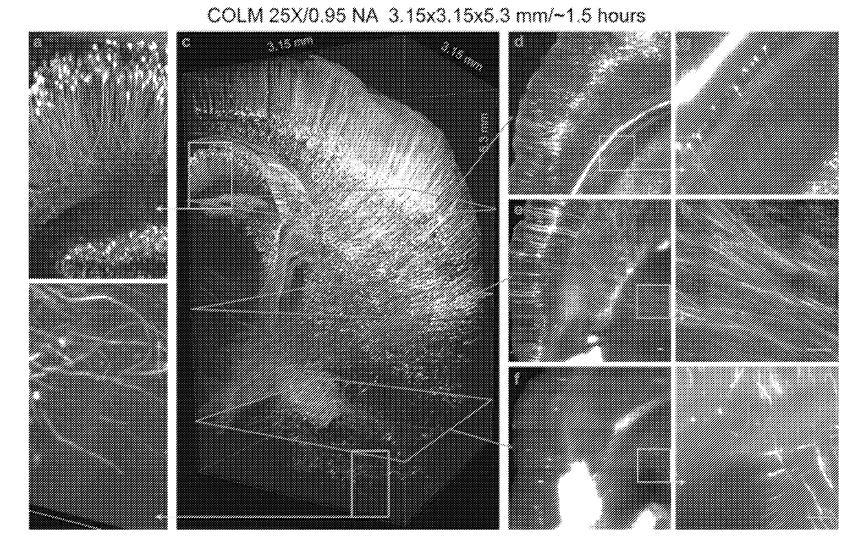
[0102]Thy1-eYFP mouse brain was perfused with 0.5% acrylamide monomer solution. A 3.15 mm×3.15 mm×5.3 mm volume was acquired from an intact clarified brain using COLM with 25× magnification. The complete image dataset was acquired in ˜1.5 hours; for optimal contrast the LUT of zoomed-in images was linearly adjusted between panels. Magnified views from FIG. 3 panel c region defined by rectangles were obtained (FIG. 3, Panels a, b). Maximum-intensity projections of over a 50 micron-thick volume were obtained (FIG. 3, Panels d-i, shown by the progression of boxes and arrows). Camera exposure time of 20 ms was used; refractive index liquid 1.454 was used as the immersion medium.

example 3

Molecular Interrogation of Clarified Tissue

[0103]Whole mouse brain was perfused with 4% acrylamide monomer solution and clarified passively, and immunostained to label all parvalbumin (PV) positive neurons. The intact brain was imaged using COLM with a 25× objective. Labeled cells at different depths in the sample were observed (FIG. 4B).

[0104]CLARITY optimized objectives were used to image clarified mouse brain tissue blocks with a confocal microscope (FIGS. 4A and C). The mouse brain was perfused with 4% acrylamide monomer solution and immunostained with anti-PV antibody. Confocal microscopy with CLARITY optimized water-immersion objectives (10×, 0.6 NA, 3 mm) was used to acquire high quality images of a 1 mm thick tissue block, and the images were processed to generate maximum intensity projection images of PV positive neurons (FIG. 4A).

[0105]It was also possible to perform multiple rounds of immunostaining and confocal imaging with CLARITY optimized water-immersion objectives (1...


PUM


Abstract

Methods and devices for conducting high-speed, high-resolution imaging of large intact tissue samples are provided. Aspects of the methods include placing a sample in an optically homogenous sample manipulation component, performing a calibration procedure to align a light sheet and a detection focal plane at a plurality of locations within the sample, and performing an imaging procedure on the sample to collect an image from each location. The collected images are reconstructed to form a three-dimensional image of the sample. Devices for carrying out the steps of the methods are also provided.

Description

CROSS REFERENCE TO RELATED APPLICATIONS[0001]This application claims priority benefit of the filing date of U.S. Provisional Patent Application Ser. No. 62 / 005,703, filed on May 30, 2014, the disclosure of which application is herein incorporated by reference in its entirety.INTRODUCTION[0002]A major challenge to the imaging of large intact tissue samples is to conduct the imaging process in a practically feasible period of time. Confocal microscopes suffer from slow imaging speeds, and can also damage the signal-emitting capabilities of a sample due to photo-bleaching before the sample is completely imaged. There is a need in the art for high-speed, high-resolution imaging methods and devices that can be used to decrease the amount of time required to image a large tissue sample while minimizing the effects of photo-bleaching on the sample. The present invention addresses these and other needs.SUMMARY[0003]Methods and devices for conducting high-speed, high-resolution imaging of la...

Claims


Application Information

Patent Timeline
IPC IPC(8): G02B21/36G02B21/00G01N33/483G02B27/00G02B21/18H04N5/225G02B21/26
CPCG02B21/367H04N5/2256G02B21/0032G02B21/26G01N33/4833G02B21/008G02B21/0076G02B21/18G02B27/0075G02B27/58G02B27/62G02B21/10H04N23/56H04N23/673
Inventor TOMER, RAJUDEISSEROTH, KARL A.
Owner THE BOARD OF TRUSTEES OF THE LELAND STANFORD JUNIOR UNIV
Why Eureka
Try Eureka