Nanocarriers for cancer treatment

a cancer and nanoparticle technology, applied in the direction of pharmaceutical delivery mechanism, organic active ingredients, radioactive preparation forms, etc., can solve the problems of reduced permeability and unclear evidence of enhanced delivery to brain tumors with small nanoparticles, and achieve the effects of increasing cerebral blood volume, increasing average vessel size, and increasing epr

Inactive Publication Date: 2017-06-15
RGT UNIV OF CALIFORNIA
View PDF0 Cites 4 Cited by
  • Summary
  • Abstract
  • Description
  • Claims
  • Application Information

AI Technical Summary

Benefits of technology

[0010]From the bio distribution data obtained after perfusion (FIG. 7), the greater accumulation associated with a greater EPR effect in an advanced xenograft (>100 mm3) was confirmed. Micelle accumulation was greater than that of liposomes regardless of the progression of the xenograft (FIG. 7a). Although the radioactivity in the left brain was ˜10-fold lower than in the right brain (FIG. 7b), the accumulation in the normal left brain showed two significant effects associated with the adjacent disease. First, in the contralateral left brain, 3HM uptake increased with xenograft progression in the implanted right brain. The permeability of the contralateral brain could be affected by the pressure induced by the growing tumor or by cytokines and growth factors associated within tumor [53]. Second, the 110-nm liposomal uptake in the left brain was similar (˜0.008% ID / g) regardless of the glioblastoma diameter. Thus, the extravasation of 110-nm liposomes was limited by the vascular pore size cutoff but relatively small 20-nm micelles crossed the BBB.
[0011]A major advantage of the PET-MRI techniques applied here is the opportunity to simultaneously estimate the PK and the local blood volume. Extended circulation of nanoparticles in the blood is crucial for the extravasation through leaky vasculature and accumulation in tumors. In our previous PK studies of liposomes and micelles in a mouse model [40, 54], the half-life of 64Cu-liposomes and 64Cu-micelles was 18 and 25 h (one-phase decay), respectively. Here, we observed a shorter half-life for both particles in blood (t1 / 2 liposomes and micelles=16.5 and 15.5 h). The observed circulation time was longer than 99mTc-labeled HYNIC-PEG liposomes previously studied in a rat model where only 52% ID remained in the blood pool 4 h after injection [55]. We assume that the reduced half-life observed here was due to differences in the vascular physiology between the two species. Here, the similar blood clearance of the nanoparticles in blood facilitated a direct comparison of the radioactivity in the tissues at the same time point.
[0012]When evaluating long-circulating nanoparticles, the blood volume can also be estimated by evaluating the radioactivity in the blood and tumor at the time of injection as calculated by a previously described radiometric method [51]. Previous MR studies in the rat brain reported a relationship between blood volume and vessel size where approximately 15% of C6 gliomas demonstrated an increased cerebral blood volume as compared to gray matter, and 90% demonstrated an increased average vessel size [50]. In a subsequent study, no correlation was found between blood vessel density and tumor progression in GBM [56]. Here, we observed a 62-82% increase in the % vascular volume in the tumor as compared to the contralateral LBV (FIG. 6a) but the % vascular volume was not significantly different between small (<100 mm3) and large tumors (>100 mm3) (FIG. 6b). The vascular volume in the adjacent left brain also was not significantly changed with xenograft progression (FIG. S4). Immunohistochemistry (IHC) with a CD31 antibody demonstrated larger vessels (FIG. 8a, black arrow) in glioblastoma lesions, which were not observed in normal brain tissue (striatum, FIG. 8b, black arrow). Previous work also demonstrated large vessels in tumors larger than 4 mm [57]. In addition, in our study MR images (arrows in FIG. 3 and FIG. 4a-b) resolved large vessels within glioblastoma lesions. The TBV and LBV results suggest that vascularization of glioblastoma increases the vascular volume in glioblastoma.
[0013]The biodistribution of both nanoparticles in organs such as the heart, lung, stomach, muscle, bone, liver and kidney was similar. As we observed in our previous study [40], the micelle accumulation was significantly lower in spleen than that observed with liposomes, which could ultimately reduce the treatment toxicity.
[0014]Recently, 3HM micelles were loaded with doxorubicin and prolonged drug bioavailability in circulation [42, 43], which may improve therapeutic efficacy and reduce splenic toxicity. Success in ongoing research with respect to loading or conjugating anticancer drugs to micelles could provide a promising method to treat glioblastoma [58, 59].
[0015]In conclusion, current GBM treatment includes invasive surgery, radiotherapy, and chemotherapy; however, drug delivery remains a major challenge. Here, we demonstrated that 3HM accumulate within glioblastoma to a significantly greater extent than 110-nm liposomes. PET / MR co-registration of brain images with multiple imaging modalities may facilitate the monitoring of disease progression and planning of treatment regimens.

Problems solved by technology

Although previous studies have demonstrated that vascular permeability is reduced in brain tumors compared to tumors within other organs, enhanced delivery to brain tumors with small nanoparticles has not been clearly demonstrated.

Method used


Image

  • Nanocarriers for cancer treatment
  • Nanocarriers for cancer treatment
  • Nanocarriers for cancer treatment

Examples


example 2

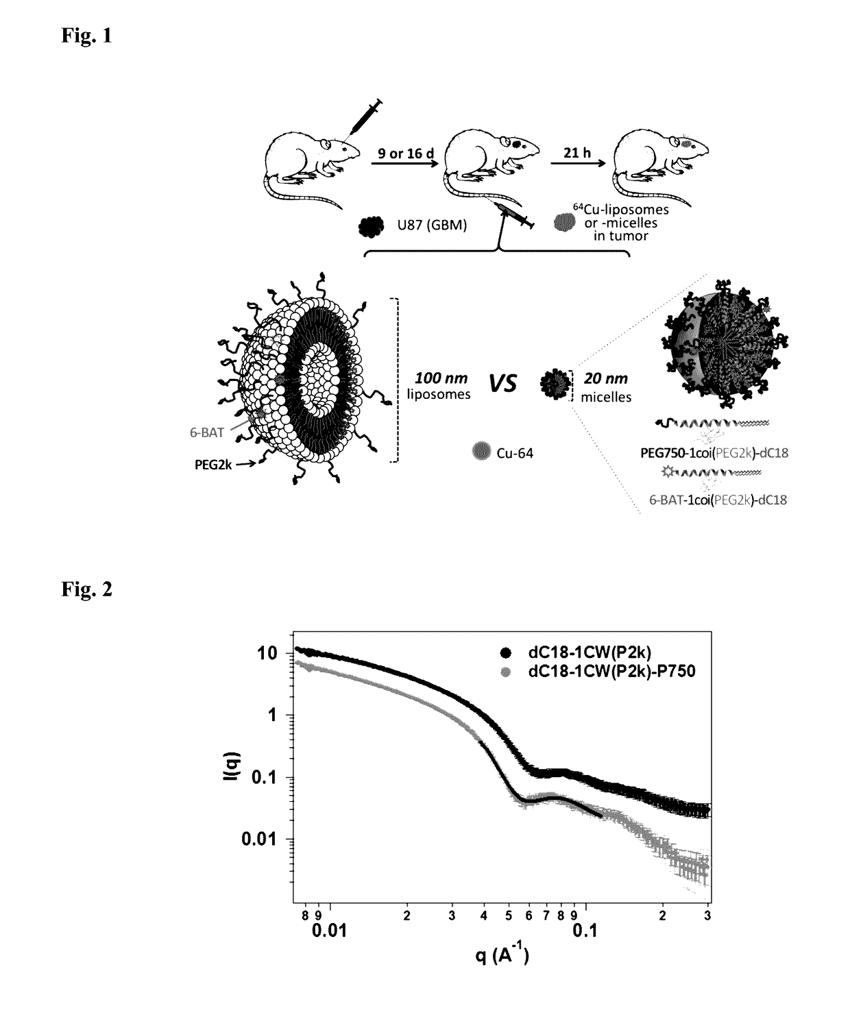
Preparation of 64Cu-Liposomes and -Micelles

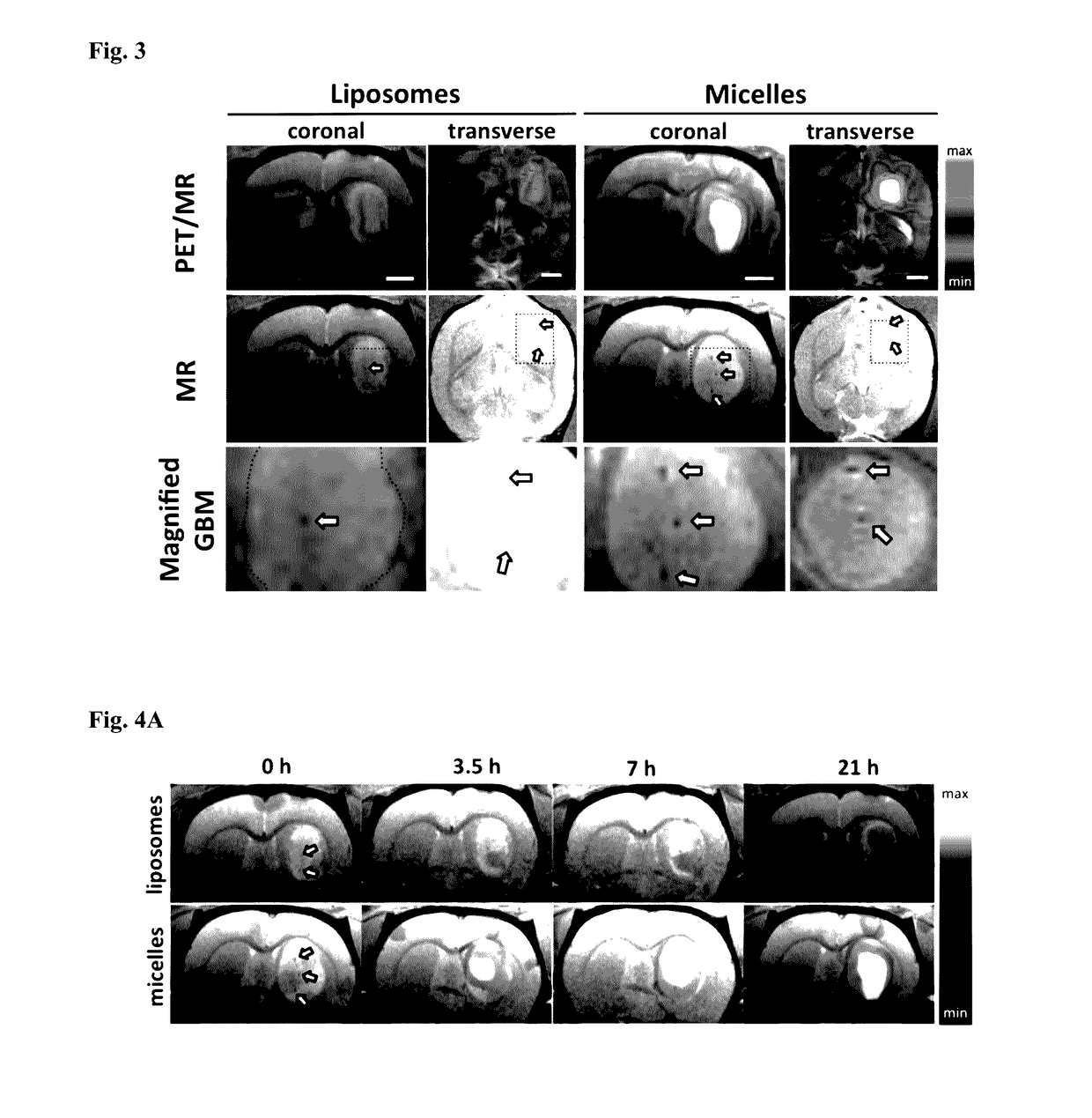
[0145]To facilitate post-labeling, a custom lipid-PEG-chelator conjugate is incorporated into the self-assembled liposomes and micelles. As illustrated in FIG. 1, liposomes with 0.5 mol % 6-BAT lipid and micelles with 2 mol % of dC18-1COI(P2k)-6-BAT were successfully prepared in 0.1 M ammonium citrate buffer (pH 5.5) and deionized water, respectively. The average mean diameter of the liposomes and micelles was 111.9±5.7 and 19.6±7.4 nm, respectively (Table 1). The Z-average particle size of the liposomes was about 6-fold greater than that of the micelles (FIG. 1). The zeta-potential of the liposomes and micelles was −15.6±3.5 and -13.6±1.4 mV under physiological pH, where the negative charge of micelles and liposomes results from PEG on the surface. 64Cu was efficiently incorporated into the 6-BAT chelator on both particles resulting in an 80±19% radiolabeling yield, which is comparable to the previous reports [39, 40]. The radiochemical pu...


PUM

PropertyMeasurementUnit
molecular weightaaaaaaaaaa
diameteraaaaaaaaaa
sizeaaaaaaaaaa

Abstract

The present invention provides conjugates containing metal binding ligands, as well as nanocarriers prepared from the conjugates.

Description

CROSS-REFERENCES TO RELATED APPLICATIONS[0001]The present application claims priority to U.S. Provisional Pat. Appl. No. 62 / 254,508, filed Nov. 12, 2015, which application is incorporated herein by reference in its entirety for all purposes.STATEMENT AS TO RIGHTS TO INVENTIONS MADE UNDER FEDERALLY SPONSORED RESEARCH AND DEVELOPMENT[0002]This invention was made with Government support under Grant Nos. NIHR01CA103828, R01CA134659, and R21EB016947. The Government has certain rights in this invention.BACKGROUND OF THE INVENTION[0003]Glioblastoma multiforme (GBM) is the most common and aggressive malignant primary brain tumor, with a median patient survival of 12-15 months [1-3]. Combining radiotherapy and post-surgical chemotherapy using cisplatin [4, 5], irinotecan [6-8], thalidomide [9, 10], or bevacizumab [11, 12] has only led to a limited improvement in survival rate [13, 14]. The blood-brain barrier (BBB) typically limits the accumulation of therapeutics within the brain and such d...

Claims


Application Information

Patent Timeline
Patent Type & Authority Applications(United States)
IPC IPC(8): A61K51/08A61K31/704A61K51/12
CPCA61K51/088A61K31/704A61K51/1234
Inventor XU, TINGFORSAYETH, JOHNFERRARA, KATHERINE
Owner RGT UNIV OF CALIFORNIA
Why Eureka
Try Eureka