Ultrasound imaging with sparse array probes

a technology of array probes and ultrasonic imaging, applied in the field of ultrasonic imaging, can solve the problems of slow frame rate, insufficient speed to capture motion in arteries, organs such as kidneys and especially the heart, or in moving joints or muscles, and achieve the effect of slow frame ra

Active Publication Date: 2017-07-27
MAUI IMAGING
View PDF11 Cites 33 Cited by
  • Summary
  • Abstract
  • Description
  • Claims
  • Application Information

AI Technical Summary

Benefits of technology

[0008]The various embodiments of systems and methods herein provide the ability to perform high resolution three-dimensional ultrasound imaging at frame rates sufficient to capture details of moving objects. Traditional scanline-based ultrasound imaging methods are limited to relatively slow frame rates due to the need to transmit and receive many scanlines to obtain a single two-dimensional plane. Extending such techniques to obtain imaging data from a complete 3D volume results in even slower frame rates due to the need to image many 2D slices.

Problems solved by technology

Traditional scanline-based ultrasound imaging methods are limited to relatively slow frame rates due to the need to transmit and receive many scanlines to obtain a single two-dimensional plane.
Extending such techniques to obtain imaging data from a complete 3D volume results in even slower frame rates due to the need to image many 2D slices.
This collection time may be adequate for non-moving tissue such as bone, liver, etc., but is not fast enough to capture motion in arteries, or organs such as kidneys and especially the heart, or in moving joints or muscles.

Method used


Image

  • Ultrasound imaging with sparse array probes
  • Ultrasound imaging with sparse array probes
  • Ultrasound imaging with sparse array probes

Examples


Embodiment Construction

[0083]The various embodiments will be described in detail with reference to the accompanying drawings. References made to particular examples and implementations are for illustrative purposes, and are not intended to limit the scope of the invention or the claims.

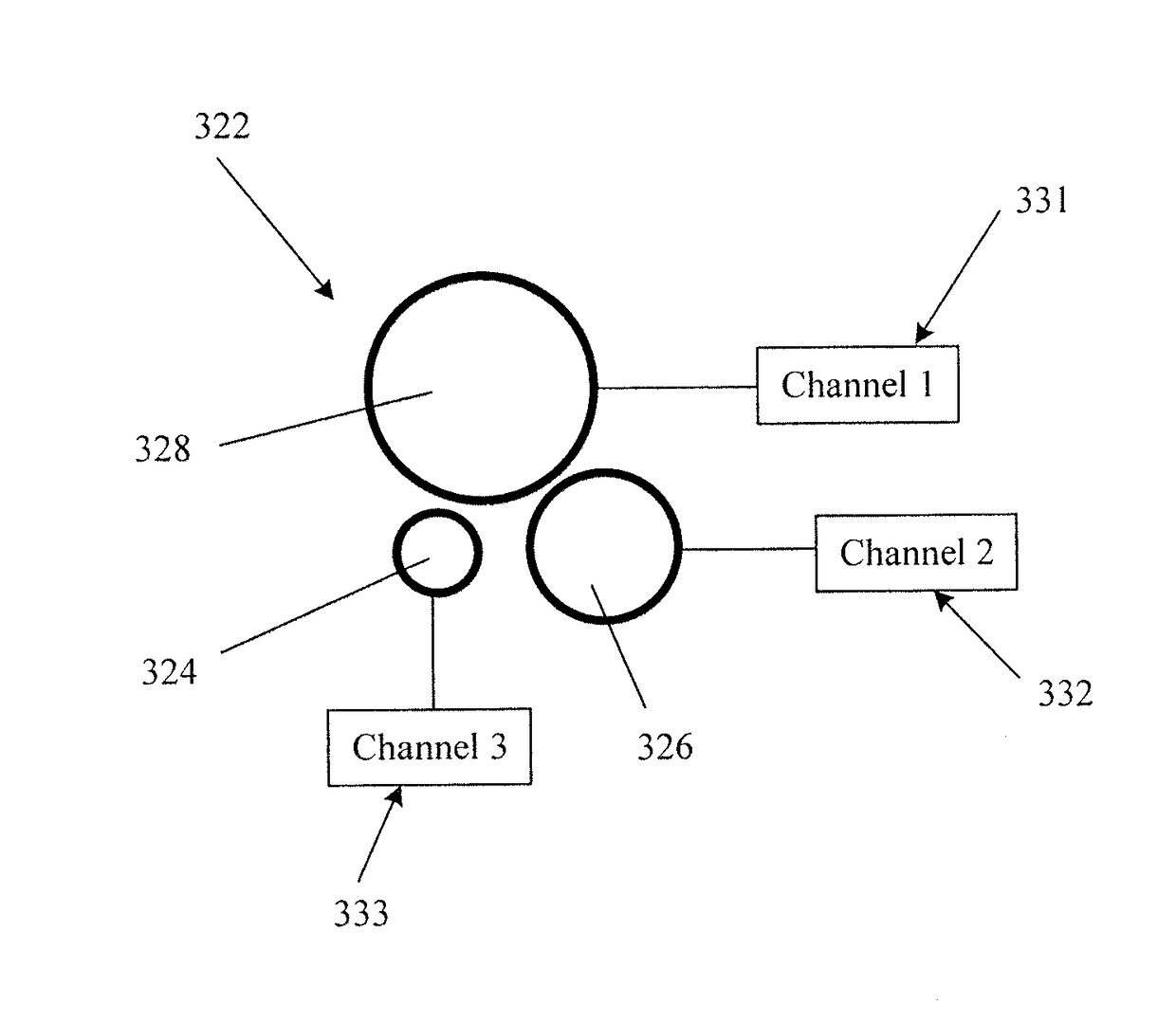
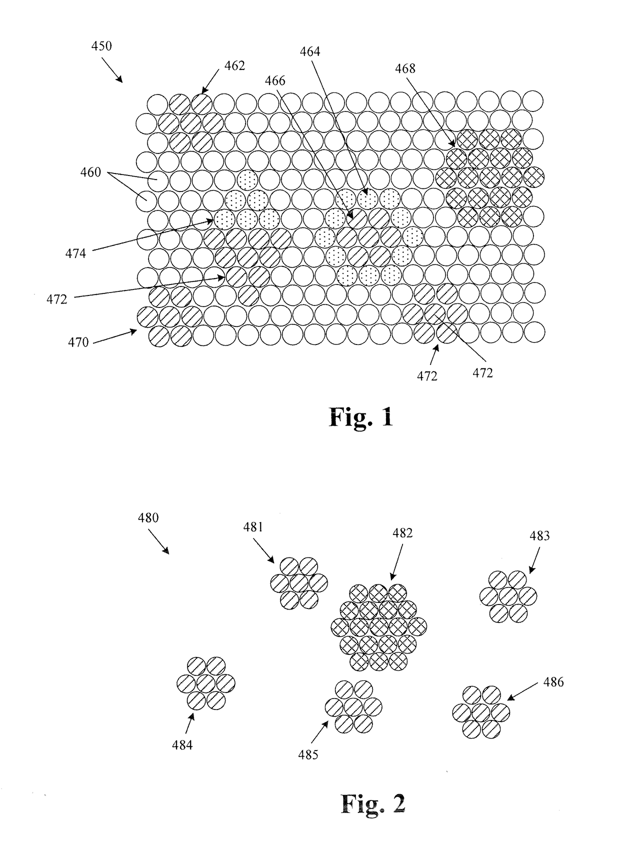
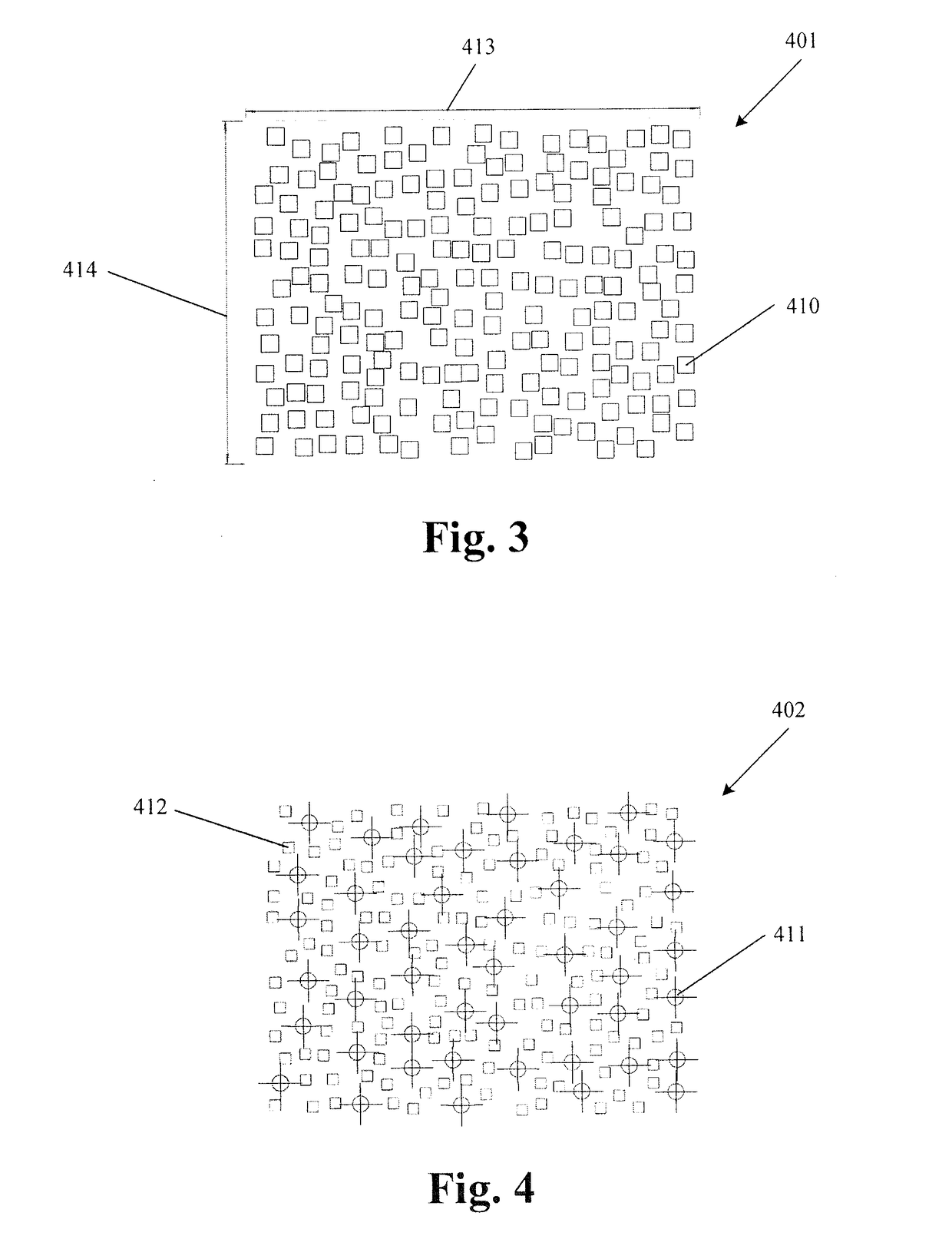
[0084]The present disclosure provides systems and methods for improving the quality of real-time two-dimensional and three-dimensional ultrasound imaging through the use of sparse arrays of various construction, including arrays in which each “element” is made up of a plurality of micro-elements arranged to be operated in concert with one another.

[0085]Although the various embodiments are described herein with reference to ultrasound imaging of various anatomic structures, it will be understood that many of the methods and devices shown and described herein may also be used in other applications, such as imaging and evaluating non-anatomic structures and objects. For example, the various embodiments herein may be applied to...


PUM


Abstract

Sparse arrays of transducer elements may be beneficial in providing ultrasound transducer probes with a wide total aperture while containing a manageable number of transducer elements. Sparse arrays made with bulk piezoelectric materials or with arrays of micro-elements can be effectively with ping-based multiple aperture ultrasound imaging techniques to perform real-time volumetric imaging.

Description

CROSS-REFERENCE TO RELATED APPLICATIONS[0001]This application claims the benefit of U.S. Provisional Patent Application No. 62 / 287,694, filed on Jan. 27, 2016, titled “Ultrasound Imaging Using Apparent Point-Source Transmit Transducer”, and U.S. Provisional Patent Application No. 62 / 310,482, filed on Mar. 18, 2016, titled “Real-Time Three-Dimensional Ultrasound Imaging” both of which are herein incorporated by reference in their entirety.INCORPORATION BY REFERENCE[0002]All publications and patent applications mentioned in this specification are herein incorporated by reference in their entirety to the same extent as if each individual publication or patent application was specifically and individually indicated to be incorporated by reference.FIELD[0003]This application relates generally to the field of ultrasound imaging, and more particularly to ping-based ultrasound imaging using sparse arrays of ultrasound transducer elements.BACKGROUND[0004]In conventional scanline-based ultras...

Claims


Application Information

Patent Timeline
Patent Type & Authority Applications(United States)
IPC IPC(8): A61B8/00A61B8/08A61B8/14
CPCA61B8/4488A61B8/483A61B8/4494A61B8/14G01S7/52079G01S15/8927G01S15/8925B06B1/0622
Inventor DAVIS, SR., HENRY A.SPECHT, DONALD F.CALL, JOSEF R.ADAM, SHARON L.SMITH, DAVID M.GOKBORA, ERIK
Owner MAUI IMAGING
Why Eureka
Try Eureka