Methods and articles for delivering viable cells into solid tissue

a technology of solid tissue and viable cells, which is applied in the direction of catheters, applications, metabolic disorders, etc., can solve the problems of slow or erratic absorption of viable cells, breakdown/degradation of viable cells, and limited application of proteins, antibodies and peptides, etc., to promote viability and enhance penetration into tissue

Active Publication Date: 2017-09-14
INCUBE LABS
View PDF1 Cites 20 Cited by
  • Summary
  • Abstract
  • Description
  • Claims
  • Application Information

AI Technical Summary

Benefits of technology

[0011]In a first aspect, the present invention provides articles for delivering viable cells into solid tissue. The articles include a shell configured for self-penetration into the solid tissue, typically having a sharpened, pointed, conical, or other conventional tissue-penetrating tip. In other embodiments, the self-penetrating tip may include an electrosurgical element (e.g. and RF energy powered element) to enhance penetration into tissue. A mass of viable therapeutic cells is maintained within an interior of the shell under conditions which promote viability. In particular embodiments, cells will usually be maintained in preparation including a viability-sustaining gel, such as an alginate gel, a protein gel, a glycosaminoglycan gel, a carbohydrate gel, or other conventional gel or medium known for maintaining mammalian cell viability. In particular embodiments the gel can be saturated or partially saturated with oxygen to provide further viability sustaining qualities to the gel. This may be done before or after addition of the cells to the gel. Once penetrated or otherwise placed into tissue, the article is configured to degrade (also referred to as biodegrade) in tissue to release the mass of viable cells into tissue.
[0012]In certain embodiments, the shell of the article will be configured to biodegrade over a time period of at least 12 hours, i.e. the shell will not degrade during the initial 12 hours it is within the patient gastrointestinal tract or elsewhere. Often, the time period for degradation is in the range from 4 hours to one year, typically being from ½ days to 5 days with specific embodiments of 1, 2, 3, 4 days. Often, it will be an objective of the present invention to maintain the shell only for so long as necessary to protect the cells from passage through the gastrointestinal tract and to release the shells from containment as quickly as possible after implantation into the intestinal wall or other location.
[0013]A variety of specific materials may be used for the biodegradable shells, including biodegradable metals, such as magnesium, iron, and zinc. Biodegradable polymers will also be useful, including, for example, poly lactic acid (PLA), poly lactic-co-glycolic acid (PGLA) and various sugars such as maltose, sucrose and the like. Further according to one or more embodiments the shell may comprise multiple layers of the same or different materials with the layers configured to degrade at different rates (e.g. hours vs days). In use such embodiments allow for a rapid release of cells followed for example for the rapid generation of a particular therapeutic substance such as insulin, followed by a longer term release to allow for the longer term release. For embodiments of the article configured for the treatment of diabetes or other glucose regulation disorder this allows for a short term and longer term treatment of the disease or condition. The variation in the degradation rates can be controlled by selection of one or more of the thickness, surface area, material and material properties of the shell.
[0014]In certain embodiments, the shell of the delivery article may have fenestrations, e.g., holes, apertures, or other small passages formed there through. The fenestrations will be configured to allow passage of fluids and small molecules into and out of an interior of the shell, but to contain the cells and the gel within the interior of the shell. In various embodiments the fenestrations can have a diameter a selected diameter matched to a selected percentage (e.g., 1, 5, 10, 25, 50% etc.) to the major diameter of the particular cell or cells contained within the article. The fenestrations will typically have a width in the range from about 0.1 to 15 μm more preferably about 0.5 to 5 μm and / or an area in the range from 0.03 to 700 μm2 more preferably about 0.8 to 80 μm2. When the shell comprises fenestrations, it may be formed from a non-biodegradable material since many cells can maintain viability through fluid and substance exchange with the implanted tissue and can release therapeutically useful cell products to the tissue through the same fenestrations. Usually, however, even with fenestrations, it will be preferable to form the wall from the biodegradable material. In an alternative or additional embodiment the fenestrations may be configured to produce a distinct acoustical signature or pattern when pinged by an external acoustical transceiver such as an ultrasound transceiver or other piezo electric based acoustical transceiver. Further as the fenestrations break down due to bioerosion of the article the acoustical signature of shell in response to the ping changes such the degree of bio-erosion of the shell can be discerned. In particular embodiments the fenestrations can be configured to produce distinct acoustical patterns when there is no bio-erosion, and when there is 25, 50 and 75% bio-erosion. In this way, the fenestrations provide actionable information for the user to know when the article has degraded sufficiently to release the cells to the selected tissue site. The specific acoustical signatures can be produced by the size and spacing of the fenestrations. In alternative or additional embodiments for providing information on the state of degradation of the articles, the articles can include acoustical markers or indicia configured to provide specific acoustical signatures of the percent degradation of the article.
[0015]In a second aspect, the present invention provides methods for delivering viable cells into solid tissues such as the tissue in the walls of the intestinal tract such as the small intestine and / or the digestive organs such as the pancreas. The methods comprise administering an article to a patient, such as a tissue penetrating member, where the article contains a selected mass or volume of therapeutic cells in a viable state. Typically, the mass of cells will be contained in preparation comprising the cells and a viability sustaining gel described herein. The article is then penetrated into the solid tissue, and at least a portion of the article biodegrades to release the cells within the tissue in a viable state. The cells are then available to produce therapeutically useful agents and substances which can be released directly into the tissue in which the cells have been implanted. In certain embodiments of this method, administering comprises the patient swallowing the article, which may be contained in swallowable capsule described herein, where the article passes through the patient's gastrointestinal tract and into the patient's intestines. Once resident in the intestines, the article will be propelled into the intestinal wall by operative coupling with an expansion means such as an expandable balloon that contains chemical reactants which produce a gas upon being mixed so to expand the balloon. Typically, this will be done response to a pH change within the intestines, such as the higher pH in the small intestine, where said pH change triggers the chemical reaction which results in propulsion of the article into the intestinal wall. Other means for generating a propulsive force so at to have the article penetrate and be inserted into the intestinal wall are also contemplated. For example, a propulsive force may be generated through the use of an electromagnetic force (from piezo electro material), a hydrostatic force or a spring force.
[0016]In a third aspect, the invention provides methods for delivering viable cells into solid tissues such as the tissue in the walls of the intestinal tract wherein the cells are put into a reversible suspended state of animation prior to being administered to the patient (including prior to being put into the article) wherein they reanimate after being delivered to the patient so as to produce a therapeutic effect. Such therapeutic effects can include for example the cellular production of one or more therapeutic compounds (e.g., insulin, integrins, etc.). Typically, this will be done by freezing or chilling the cells and / or the gel containing them within the article prior to administering the article to the patient. Chilling may done to a temperature in the range of 50 to 33° F. with a preferred range of 39 to 40° F. In an alternative or variation, the cells and / or gel containing them may be frozen or chilled prior to being placed into the article. Freezing or chilling the articles and the cells therein prior to administration is particularly useful as it can preserve the cells for extended periods of time in a reversible state of suspended animation without the need to supply nutrients and remove cellular products. Once articles are administered to the patient, however, the cells will thaw and warm within a short time period to become reanimated and will resume cellular processes and may be at risk of degradation from the gastrointestinal environment. The shell of the article, as described above, will protect the now metabolically active cells as they pass through a patient's gastrointestinal tract where they would be exposed to the digestive conditions of the stomach which, without protection, would damage or kill the cells. Once penetrated into the intestinal tract, the article will be implanted into the intestinal tissue, thus releasing the viable cells where they are now in an environment in which cell viability can be maintained and they can generate the useful therapeutic substances which they produce. Embodiments of these methods employ articles which may have the preferred characteristics and dimensions discussed above in connection with articles of the present invention. Other methods of suspended reversible animation of the viable cells are also contemplated besides freezing and chilling. These may include for example, use of certain compounds in the gel and / or charging the gel with an inert gas (e.g., nitrogen) and lyophilizing of the cells (or other freeze drying methods) prior to administration.

Problems solved by technology

While there has been an increasing development of new viable cells in recent years for the treatment of a variety of diseases, many including proteins, antibodies and peptides have limited application because they cannot be given orally.
This is due to a number of reasons including: poor oral toleration with complications including gastric irritation and bleeding; breakdown / degradation of the viable cells compounds in the stomach; and poor, slow or erratic absorption of the viable cells.
Conventional alternative viable cells delivery methods such as intravenous and intramuscular delivery have a number of drawbacks including pain and risk of infection from a needle stick, requirements for the use of sterile technique and the requirement and associated risks of maintaining an IV line in a patient for an extended period of time.
While other viable cells delivery approaches have been employed such as implantable viable cells delivery pumps, these approaches require the semi-permanent implantation of a device and can still have many of the limitations of IV delivery.
While very effective for delivering viable cells formulations, these devices are not optimized for delivering living cells to a patient.
The delivery of living or viable cells, such as stem cells, cells which secrete therapeutically beneficial agents, and the like, to a patient promises to be of enormous clinical benefit, but is hindered by a shortage of effective delivery apparatus, materials and protocols.

Method used


Image

  • Methods and articles for delivering viable cells into solid tissue
  • Methods and articles for delivering viable cells into solid tissue
  • Methods and articles for delivering viable cells into solid tissue

Examples


Embodiment Construction

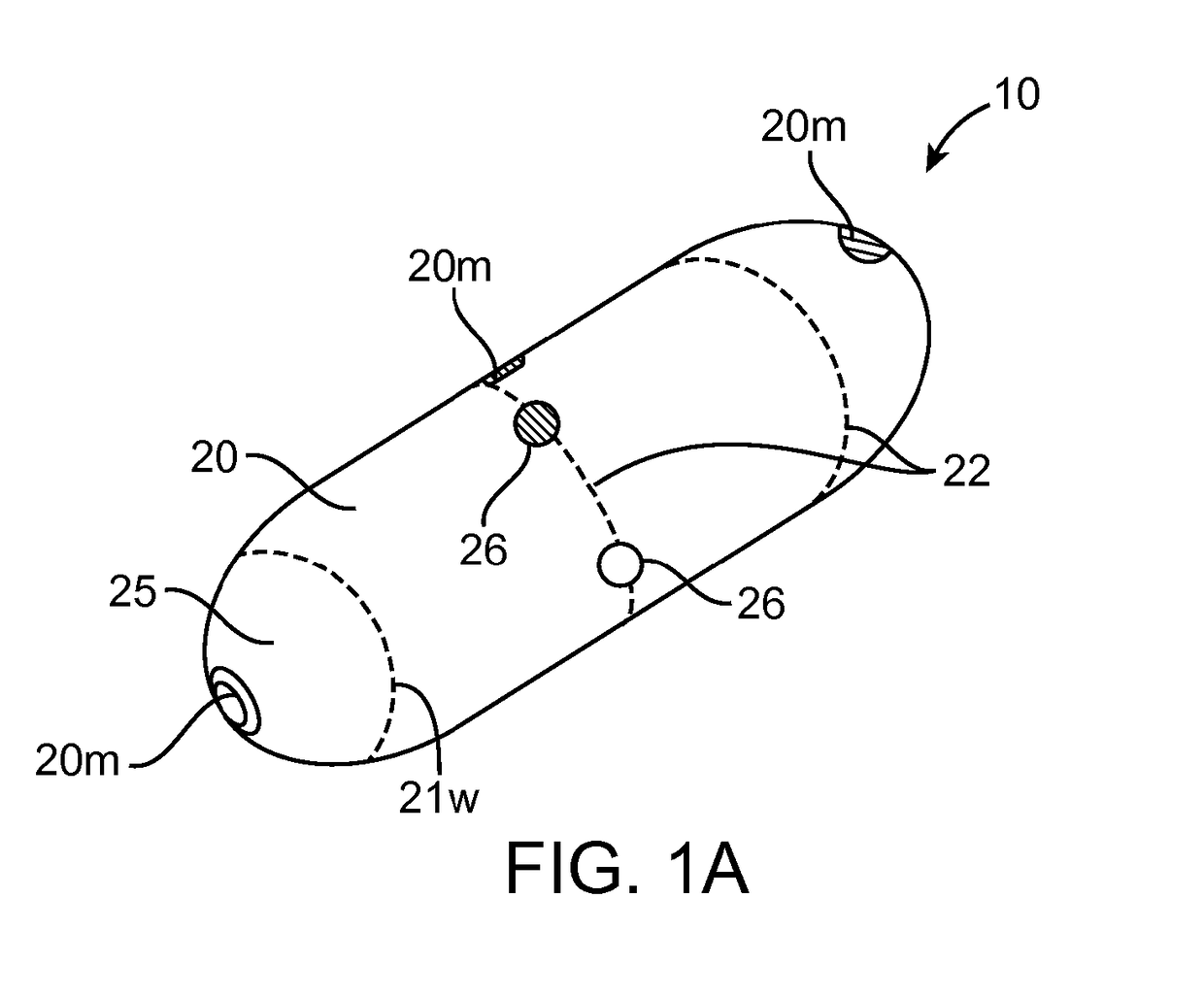
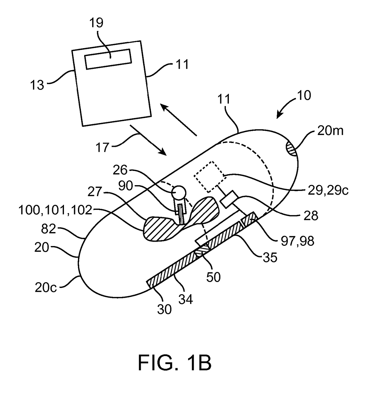
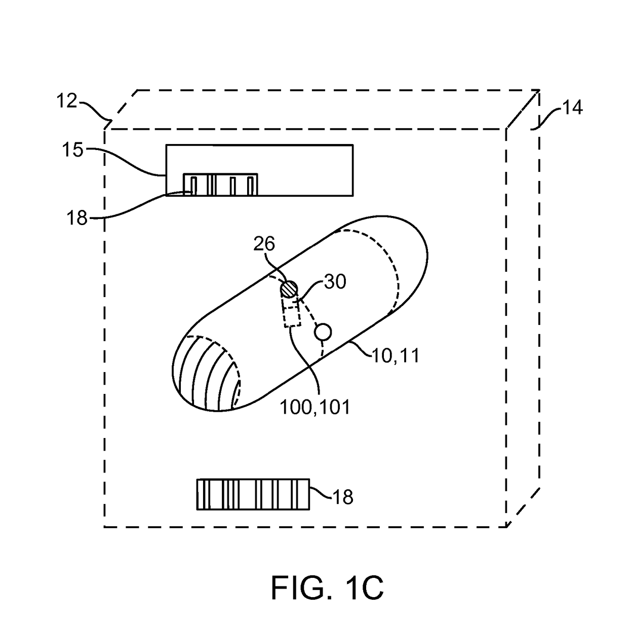
[0049]Embodiments of the invention provide devices, preparations, systems and methods for delivering viable cells in to various locations in the body, in particular to the intestinal wall tissue. In many embodiments the cells will be delivered by a swallowable device configured to maintain viability of the cells while they pass through the gastrointestinal tract and to a selected site within intestinal tract or other location. As used herein, “GI tract” refers to the esophagus, stomach, small intestine, large intestine and anus, while “Intestinal tract” refers to the small and large intestine. Various embodiments of the invention can be configured and arranged for delivery of viable cells into the intestinal tract as well as the entire GI tract. Also in many embodiments the delivered cells will comprise therapeutic cells and will sometimes be referred to as such. However embodiments of the invention are not limited to therapeutic cells only. In particular it should be understood tha...


PUM


Abstract

Embodiments provide swallowable devices, preparations and methods for delivering viable cells (VC) into the GI tract including GI wall tissue or other tissue site. Particular embodiments provide a swallowable device such as a capsule for delivering VC into an intestinal wall or other site. The VC can be contained within a tissue-penetrating shell disposed in the capsule that protects the VC as they pass through the GI tract until they are inserted into GI tract tissue or other location. The shell desirably has shape, size and material consistency to be contained in a swallowable capsule, delivered from the capsule into solid tissue by the application of force on the shell and biodegrade within the solid tissue to release the VC into the tissue. Within the shell or other structure the VC can be maintained in a viability-sustaining gel that preserves the viability of the VC for selected time periods.

Description

CROSS-REFERENCE TO RELATED APPLICATIONS[0001]This application claims the benefit of priority to Provisional U.S. Patent Application No. 62 / 305,878, entitled “Methods And Articles For Delivering Viable Cells Into Solid Tissue”, filed Mar. 9, 2016; the entire content of which is incorporated herein by reference.[0002]This application is also related to co-filed U.S. patent application Ser. No. ______ (Attorney Docket No. 42197-737.201), entitled “Methods And Articles For Delivering Viable Cells Into Solid Tissue”, the full disclosure of which is incorporated herein for all purposes. This disclosure of the application incorporates disclosure from U.S. patent application Ser. No. 13 / 837,025 (Attorney Docket No. 42197-715.501, now U.S. Pat. No. 8,734,429), filed Mar. 15, 2013, the entire contents of which are hereby incorporated by reference herein for all purposes.BACKGROUND OF THE INVENTION[0003]1. Field of the Invention[0004]Embodiments of the invention relate to swallowable viable ce...

Claims


Application Information

Patent Timeline
Patent Type & Authority Applications(United States)
IPC IPC(8): A61K9/48A61K9/00A61K35/12
CPCA61K9/4816A61K9/0053A61K35/12A61B5/6861A61B5/14507A61B5/14539A61B5/14546A61B5/4255A61M31/002A61K35/28A61K35/30A61K35/39A61K9/4891A61P3/10A61P43/00A61P5/00A61P5/48A61P5/50A61K9/06A61M25/1002A61M2025/105A61M2205/3633
Inventor IMRAN, MIRHARRIS, JOEL
Owner INCUBE LABS
Why Eureka
Try Eureka