Eureka AIR delivers breakthrough ideas for toughest innovation challenges, trusted by R&D personnel around the world.

Determining a remaining time during medical imaging

Active Publication Date: 2018-01-18
SIEMENS HEALTHCARE GMBH
View PDF7 Cites 3 Cited by
  • Summary
  • Abstract
  • Description
  • Claims
  • Application Information

AI Technical Summary

Benefits of technology

The patent describes a method for monitoring a critical event during a measurement for medical imaging, based on the comparison of the current remaining time with a minimum remaining time. A warning message is output if the current remaining time is shorter than the minimum. The method also allows for changing a pulse sequence, which may influence the critical event to increase its duration. The computer program product provides a computer software code that can be executed by a computer evaluation unit, allowing for efficient and repeatable execution of the method.

Problems solved by technology

The gradient pulses may give rise to undesirable peripheral nerve stimulation (PNS) in the body of the patient.
The absorption of the HF energy may result in a heating of the bodily tissue so that burns may occur in the case of inadmissibly high local concentration of the HF energy.

Method used


Image

  • Determining a remaining time during medical imaging
  • Determining a remaining time during medical imaging
  • Determining a remaining time during medical imaging

Examples


Embodiment Construction

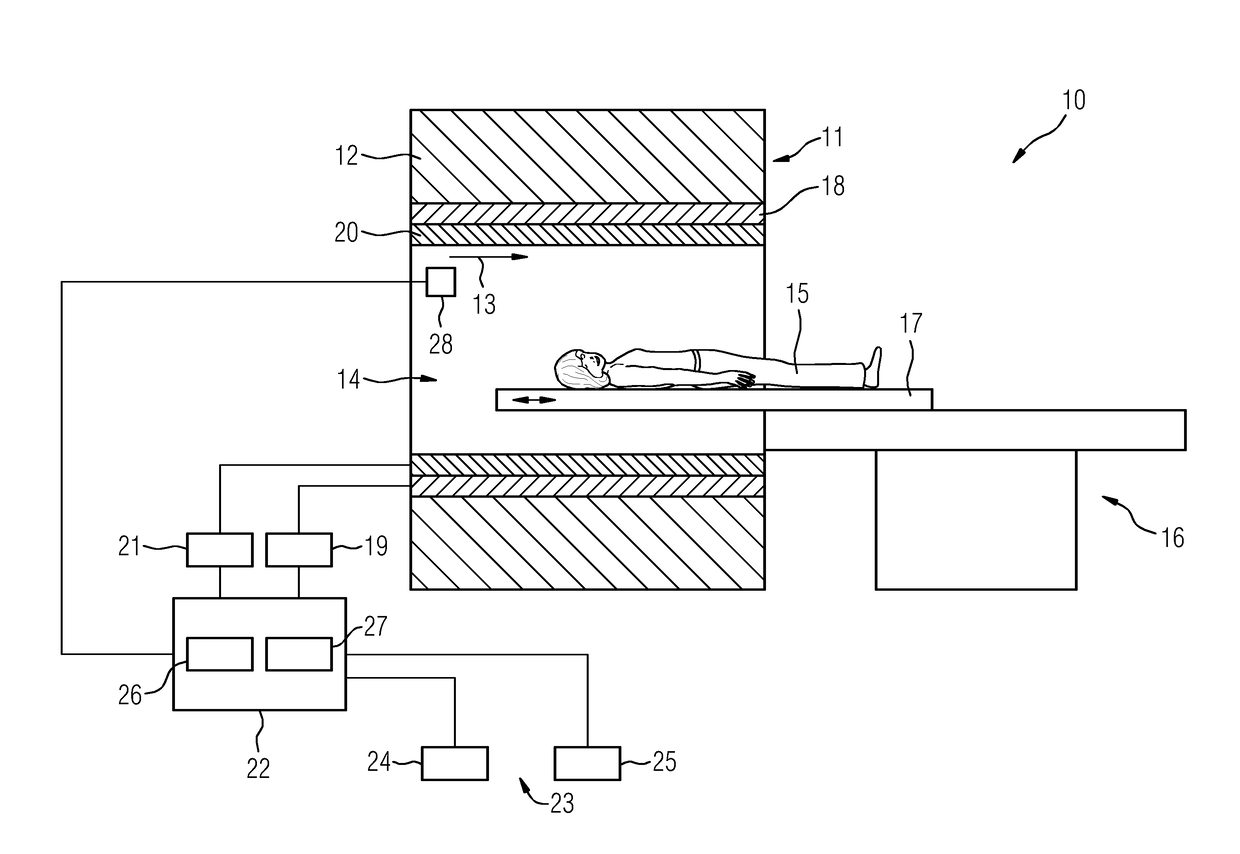
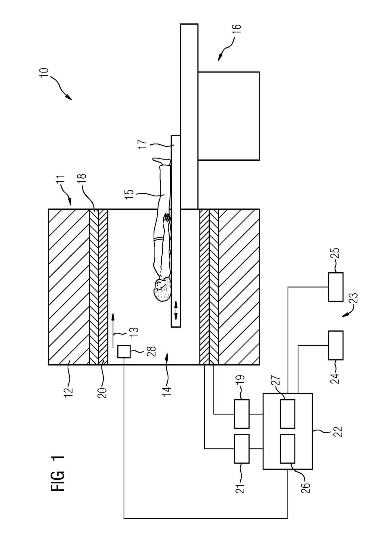
[0065]FIG. 1 shows a schematic representation of a magnetic resonance device 10 by way of example for a medical imaging device. In place of the magnetic resonance device, however, other modalities may also be employed such as, for example, a computer tomography device, an X-ray device, a mammography device, a positron emission tomography device, a single photon emission computer tomography device, a scintigraphy device, a sonography device, a thermography device, an electrical impedance tomography device, or any combination thereof.

[0066]The magnetic resonance device 10 includes a magnet unit 11 having a main magnet 12 for a generation of a main magnetic field 13 that is strong and, for example, constant over time. Additionally, the magnetic resonance device 10 includes a patient recording zone 14 for a recording of a patient 15. In the present exemplary embodiment, the patient recording zone 14 is realized in a cylindrical manner and is surrounded in a peripheral direction by the m...


PUM


Abstract

A method for determining a current remaining time during a measurement for the purpose of medical imaging, an evaluation unit, a medical imaging device, and a computer program product are provided. The method provides for a permitted operating range and an item of extrapolation information regarding the critical event to be provided. The current remaining time is determined by an evaluation unit based on the permitted operating range and the item of extrapolation information.

Description

[0001]This application claims the benefit of DE 10 2016 212 877.9, filed on Jul. 14, 2016, which is hereby incorporated by reference in its entirety.BACKGROUND[0002]The present embodiments relate to determining a current remaining time during a measurement for the purpose of medical imaging.[0003]By using medical imaging, and, for example, by using magnetic resonance tomography (MRT) and / or computed tomography (CT) and / or X-ray photographs, the interior of a body of a patient may be mapped. To do this, in the case of magnetic resonance tomography, for example, rapidly switched gradient pulses are superimposed on a static basic magnetic field during a magnetic resonance measurement with a magnetic resonance device. The gradient pulses may give rise to undesirable peripheral nerve stimulation (PNS) in the body of the patient.[0004]High-frequency excitation pulses (HF pulses) are beamed into the object under examination in order to trigger magnetic resonance signals. The energy of the ...

Claims


Application Information

Patent Timeline
IPC IPC(8): A61B6/00A61B6/03G06F19/00A61B5/055G06F3/0484
CPCA61B6/465A61B6/032A61B5/055A61B6/037G06F3/0484G06F19/3406G16H20/40G01R33/288G16H40/63
Inventor SCHNEIDER, RAINER
Owner SIEMENS HEALTHCARE GMBH
Why Eureka
Eureka Blog
Learn More