Medical Image Segmentation Method and Apparatus

Active Publication Date: 2018-04-26
FENG YUAN +1
View PDF0 Cites 20 Cited by
  • Summary
  • Abstract
  • Description
  • Claims
  • Application Information

AI Technical Summary

Benefits of technology

[0038]1. By adopting a fuzzy clustering method to segment medical images, the method can utilize the gray scale image information and the contrast value while taking into account of the balance of computation efficiency and performance. Therefore, the method can be used to segment all kinds of soft tissues and organs with low contrast and blurry boundaries. Besides, by defining the pixel difference using Hausdorff distance, the method can use the neighbor information of each pixel, especially those with low contrast, to segment the blurry boundaries.
[0039]2. The present invention can com

Problems solved by technology

However, because of the variety and complexity of the medical images, image segmentation poses a great challenge.
However, when there are many d

Method used


Image

  • Medical Image Segmentation Method and Apparatus
  • Medical Image Segmentation Method and Apparatus
  • Medical Image Segmentation Method and Apparatus

Examples


Embodiment Construction

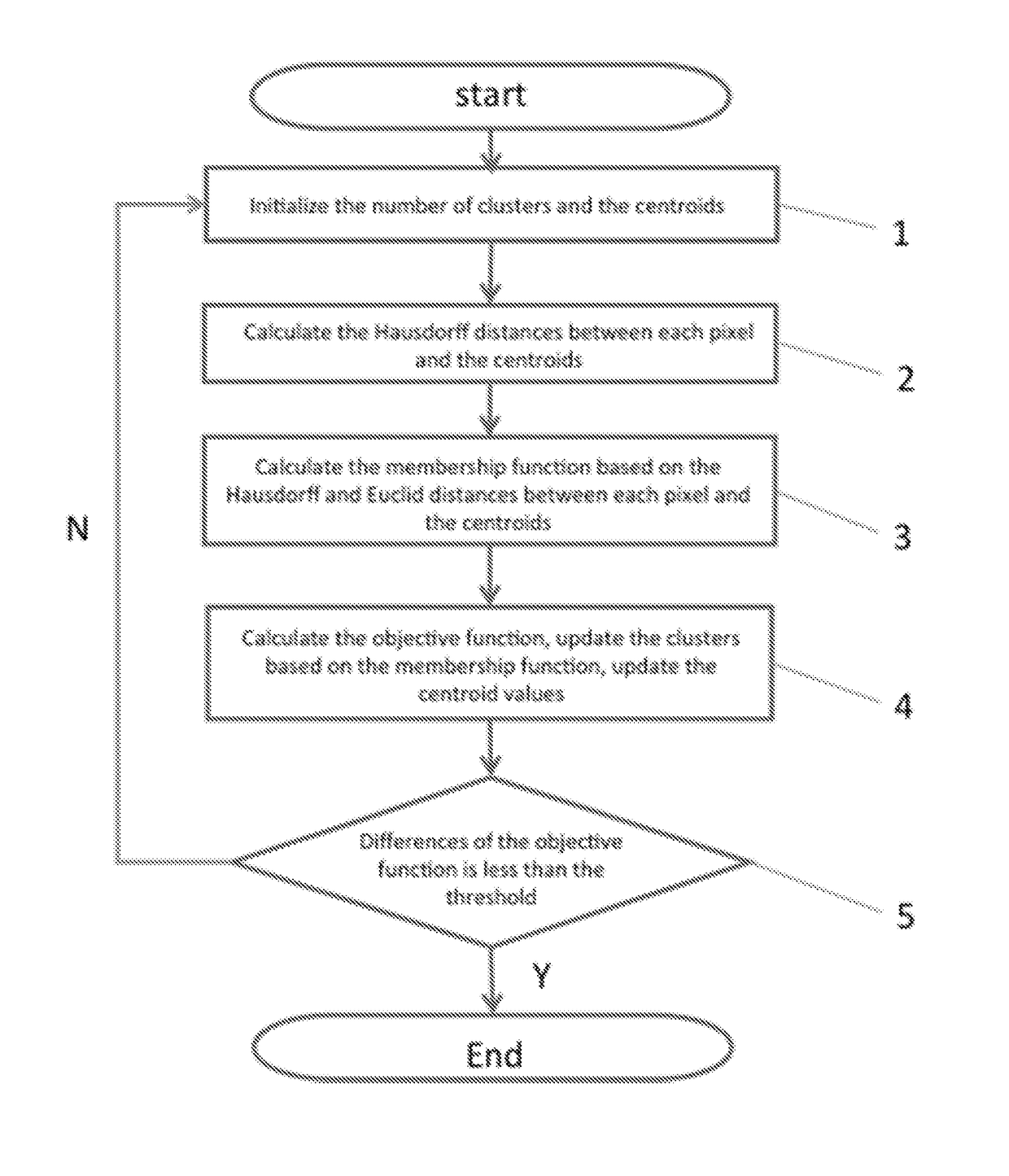
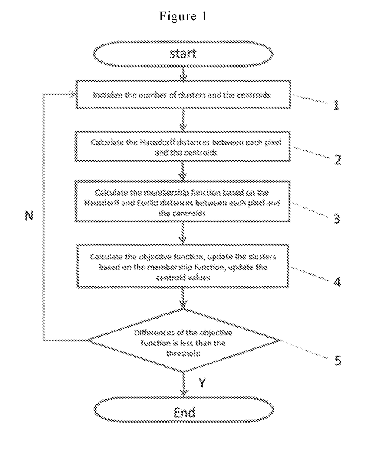
[0046]The invention will be further illustrated in more detail with reference to accompanying drawings. It is noted that, the following embodiments are intended for purposes of illustration only and are not intended to limit the scope of the invention. Any other embodiments based on the present inventions without other innovative work are also within the protection of the present invention.

[0047]In medical image research and application, only parts of the image or certain regions are of interest to scientists or clinicians. These usually include certain organs or tissues. In order to distinguish the objects for analysis, we need to extract the regions of interest out of the image for further analysis and processing. Image segmentation is a process to extract the regions of interest with different features out of an image. The features could be the gray scale level, color, or the textures of an image. The regions of interest could be a single region or different regions.

[0048]However...


PUM


Abstract

A medical image segmentation method includes: step 1, initialize of the number of clusters and determine the initial values of the cluster centroids; step 2, calculate a Hausdorff distance between each cluster centroid and each pixel in the image; step 3, calculate a membership function of each pixel based on the Hausdorff distance and a Euclid distance between the cluster centroids and each pixel; step 4, calculate an objective function, cluster the pixels of the image based on the updated membership function, and update the centroid value; and repeating step 2-4, until a difference between two objective function values is less than a threshold value. Then the membership function from the last iteration is a final segmentation.

Description

FIELD OF THE INVENTION[0001]The field of this invention is image processing. More particularly, the invention relates to a medical image segmentation method and apparatus.BACKGROUND[0002]As the development of medical imaging technology, image processing methods have been widely used in medical research and clinical applications, such as radiotherapy planning, intervention treatment, and surgical guidance. Image segmentation is one of the important topic in medical image processing. It can help the clinician to segment and extract the region of interest (pathological tissues etc.) for further analyzing and quantification, which can increase the accuracy and efficiency of clinical diagnosis. However, because of the variety and complexity of the medical images, image segmentation poses a great challenge.[0003]Among the image segmentation method used clinically, thresholding method is the most widely used because of its simple implementation and small computational load. Its algorithm i...

Claims


Application Information

Patent Timeline
IPC IPC(8): G06T7/00G06T7/11G06K9/62G06K9/52G06K9/46A61B5/00A61B6/00
CPCG06T7/0012G06T7/11G06K9/6218G06K9/52G06K2009/4666G06K9/4642A61B5/7264A61B6/5217G06K9/6215G06T2207/20081G06T2207/30024G06T2207/10072G06T2207/30004G06T7/143A61B6/5211G16H50/20G06V10/763G06F18/23213G06F18/22G06F18/23
Inventor FENG, YUANHUANG, LONG
Owner FENG YUAN
Why Eureka
Try Eureka