Image Enhancement System for Bone Disease Evaluation

Active Publication Date: 2018-11-15
WISCONSIN ALUMNI RES FOUND
View PDF0 Cites 1 Cited by
  • Summary
  • Abstract
  • Description
  • Claims
  • Application Information

AI Technical Summary

Benefits of technology

The present invention is an image enhancement system for bone disease diagnosis that corrects images to account for normal variations in different bones with respect to the uptake of a specific tracer or specific image modality. The result is a normalized image that better reveals disease location, reducing subjective interpretation. The invention uses a standard skeleton template to accentuate image features related to disease. The system statistically selects to best thresholds to identify lesions in different skeletal regions by analyzing multiple "healthy" individuals with respect to radioactive tracer uptake. The result is an improved image that may be more easily analyzed by the physician having had standard variations removed so that only disease-based differences are evident. This improved image can be used for improved automatic analysis of lesion size, location, and change.

Problems solved by technology

In analyzing lesions in the skeleton, this process is complicated by the fact that different hones have different background intensities independent of disease.
This inability to rely directly on the image for the identification of disease adds to the burden of the physician in reading these images potentially increasing the opportunity for error.
In addition, these image variations may interfere with quantitative measurements applied to the image.

Method used


Image

  • Image Enhancement System for Bone Disease Evaluation
  • Image Enhancement System for Bone Disease Evaluation
  • Image Enhancement System for Bone Disease Evaluation

Examples


Embodiment Construction

System Hardware

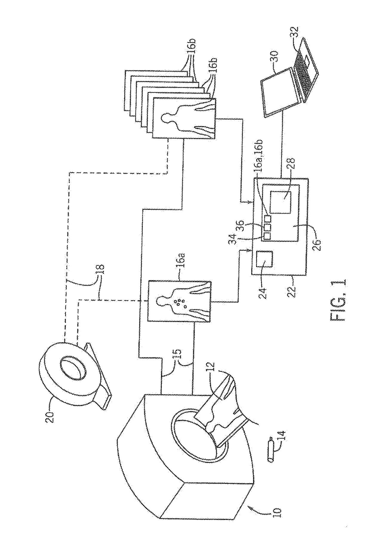
[0033]Referring now to FIG. 1, scanner 10 capable of imaging bone disease (for example, “functional imaging” or “metabolic imaging”) may scan a patient 12 after an administration to the patient 12 of a molecular imaging agent 14 (e.g., a radioactive tracer). In one embodiment, the molecular imaging agent 14 may be 18F—NaF.

[0034]The scanner 10, in one example, may be a PET (positron emission tomography) scanner. As is generally understood in the art, PET is a nuclear medical imaging technique producing three-dimensional image data comprised of multiple voxels having values revealing functional processes in the body reflected by uptake of the molecular imaging agent 14 to tumor tissue. The molecular imaging agent 14, in this case, may be a positron emitting radionuclide attached to a biologically active molecule; the latter selected to participate in the tumor's metabolism.

[0035]The patient 12 may be scanned at multiple times to produce molecular imaging data 15 providi...


PUM


Abstract

A standardized skeleton template is used to normalize medical image data of the skeleton to eliminate variations in the medical image data related to physiological variations in a normal patient thereby better accentuating disease conditions.

Description

STATEMENT REGARDING FEDERALLY SPONSORED RESEARCH OR DEVELOPMENTCROSS REFERENCE TO RELATED APPLICATIONBACKGROUND OF THE INVENTION[0001]The present invention relates to medical imaging systems, and in particular, to an imaging system enhancing bone images by removing normal variations based on standardized bone templates.[0002]In the identification of skeletal diseases, a patient may be imaged, for example using: positron emission tomography (PET), computed tomography (CT), magnetic resonance imaging (MRI), or single photon emission tomography (SPECT) to detect the abnormal image intensity and hence the presence bone disease.[0003]In the identification of some skeletal diseases, such as metastasized caner lesions, a patient may be given a radioactive tracer preferentially localizing in lesions. An image of the tracer, such as 18F—NaF, may be taken casing an imaging system such as PET / CT imaging, to detect the increased presence of the tracer and hence the presence of disease in differ...

Claims


Application Information

Patent Timeline
IPC IPC(8): G06T7/00
CPCG06T7/0012G06T2207/30096G06T2207/30008G06T2207/10072G06T2207/20012G06T5/94
Inventor JERAJ, ROBERTBRADSHAW, TYLERPERK, TIMOTHY
Owner WISCONSIN ALUMNI RES FOUND
Why Eureka
Try Eureka