
Pseudo h&e image producing method and optical system using same

a technology of h&e image and optical system, which is applied in the field of hybrid image generation of optical system, can solve the problems of damage to tissue structure, frozen cells are generally not easily stained, and it takes a lot of time for a pathologist to examine a frozen section, and achieve excellent color contrast

Active Publication Date: 2019-01-24
ACUSOLUTIONS INC
View PDF5 Cites 1 Cited by
  • Summary
  • Abstract
  • Description
  • Claims
  • Application Information

AI Technical Summary

Benefits of technology

The present invention is about a method for producing a pseudo H&E image which uses less fluorescent agent and minimizes damage to a pathological sample. It also uses less fluorescent agent and shortens the staining time to speed up the process. Additionally, it provides more detailed image information of the cytoplasm and nuclei, improving the distinguishability between normal and abnormal tissues.

Problems solved by technology

In conventional tumor surgery, it takes a lot of time for a pathologist to examine a frozen section to determine if a tumor is cleanly removed.
However, during the process of preparing a frozen section of a pathological sample, if the pathological sample contains much water, a formed crystal ice structure will cause a damage to the tissue structure of the pathological sample; and if the pathological sample contains much fat, the fat will remain unfrozen at temperatures around −20° C. where the other tissues are already frozen, and is therefore easy to slip off from the frozen section to cause an artifact to the frozen section.
Besides, frozen cells are generally not easily stained.
However, when a person has viewed a lot of red-and-green images with black background or has viewed a red-and-green image with black background for a long time, his / her eyes will feel uncomfortable and fatigued.
As a result, the format of red-and-green image with black background is not suitable for the image of a pathological sample, because a pathologist may have to inspect a lot of images of pathological samples during a work day.
However, as traditional pathological image producing method not only has an artifact issue, but also requires a long time, it is therefore not suitable for providing a timely pathological image needed in a surgery.
However, as the U.S. Pat. No. 8,269,827B2 uses fluorescent images to generate a pseudo H&E image, it has disadvantages as follows: (1) the color contrast of the pseudo H&E image is not good enough due to a fact that the internal structure of a pathological sample cannot be uniformly stained with a fluorescent agent; (2) the fluorescent agents used for generating the fluorescent images will cause damage to the pathological sample; and (3) it takes a lot of time to stain the fluorescent agents onto the pathological sample.

Method used


Image

  • Pseudo h&e image producing method and optical system using same
  • Pseudo h&e image producing method and optical system using same
  • Pseudo h&e image producing method and optical system using same

Examples


Embodiment Construction

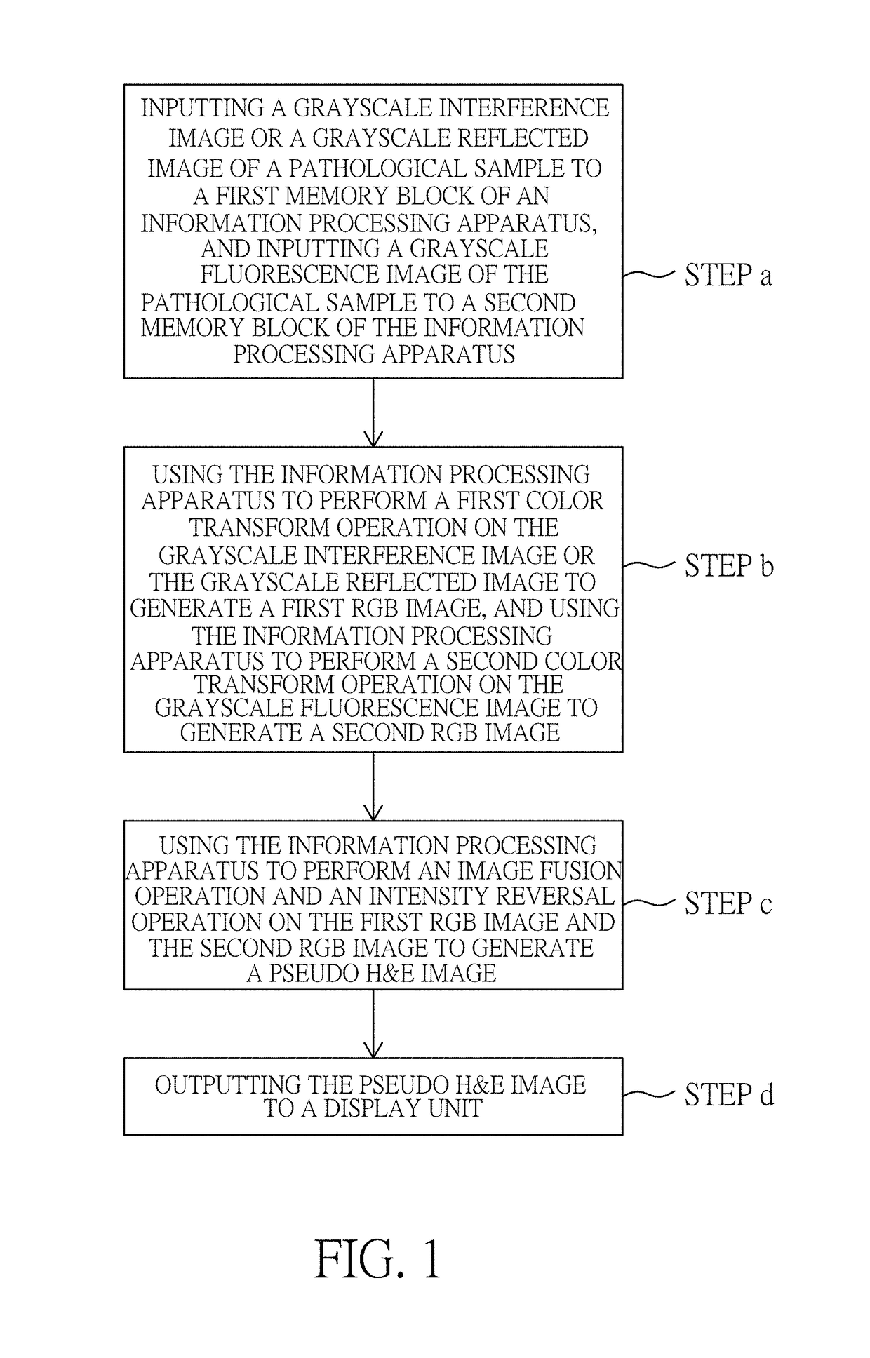
[0033]Please refer to FIG. 1, which illustrates a flow chart of pseudo H&E image producing method according to a preferred embodiment of the present invention.

[0034]As illustrated in FIG. 1, the pseudo H&E image producing method of the present invention includes the steps of:

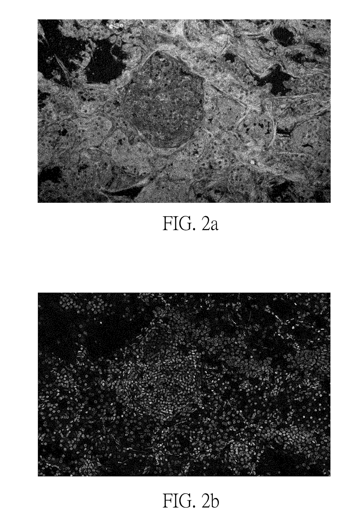
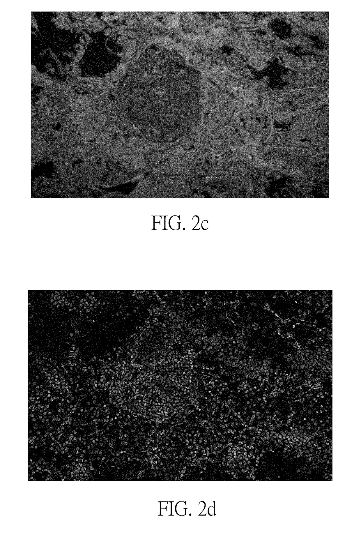
[0035]inputting a grayscale interference image or a grayscale reflected image of a pathological sample to a first memory block of an information processing apparatus, and inputting a grayscale fluorescence image of the pathological sample to a second memory block of the information processing apparatus (step a); using the information processing apparatus to perform a first color transform operation on the grayscale interference image or the grayscale reflected image to generate a first RGB image, and using the information processing apparatus to perform a second color transform operation on the grayscale fluorescence image to generate a second RGB image (step b); using the information processing apparatus to per...


PUM


Abstract

A pseudo H&E image producing method, including: inputting a grayscale interference image or a grayscale reflected image of a pathological sample to a first memory block of an information processing apparatus, and inputting a grayscale fluorescence image of the pathological sample to a second memory block of the information processing apparatus; using the information processing apparatus to perform a first color transform operation on the grayscale interference image or the grayscale reflected image to generate a first RGB image, and using the information processing apparatus to perform a second color transform operation on the grayscale fluorescence image to generate a second RGB image; and using the information processing apparatus to perform an image fusion operation and an intensity reversal operation on the first RGB image and the second RGB image to generate a pseudo H&E image. The present invention also discloses an optical system using the method.

Description

BACKGROUND OF THE INVENTIONField of the Invention[0001]The present invention relates to a hybrid image generating method of an optical system, especially to a pseudo H&E image producing method and an optical system using the method.Description of the Related Art[0002]In conventional tumor surgery, it takes a lot of time for a pathologist to examine a frozen section to determine if a tumor is cleanly removed. However, during the process of preparing a frozen section of a pathological sample, if the pathological sample contains much water, a formed crystal ice structure will cause a damage to the tissue structure of the pathological sample; and if the pathological sample contains much fat, the fat will remain unfrozen at temperatures around −20° C. where the other tissues are already frozen, and is therefore easy to slip off from the frozen section to cause an artifact to the frozen section. Besides, frozen cells are generally not easily stained. Due to the facts disclosed above, the ...

Claims


Application Information

Patent Timeline
Patent Type & Authority Applications(United States)
IPC IPC(8): H04N9/43G06T7/00G06T5/50
CPCG01N21/64G01N21/6458G06T11/001G06T2207/30004G06T2207/10056G06T2207/20221G06T5/50G06T7/0014G06T2207/10024G06T2207/10064G06T2207/30024H04N9/43
Inventor TSAI, CHIEN-CHUNGHSU, KUANG-YU
Owner ACUSOLUTIONS INC
Learn More