Image synthesis method for biological sample and optical system using the same

A technology for biological sample and image synthesis, which is used in material analysis by optical means, scientific instruments, fluorescence/phosphorescence, etc. It can solve the problems of H&E-like image color contrast that needs to be improved, sample tissue damage, etc.

Pending Publication Date: 2019-10-18
ACUSOLUTIONS INC
View PDF1 Cites 0 Cited by
  • Summary
  • Abstract
  • Description
  • Claims
  • Application Information

AI Technical Summary

Problems solved by technology

[0011] However, in this patent framework, multiple fluorescent images are added through color addition to generate an H&E-like image. However (1) the fluorescent agent

Method used


Image

  • Image synthesis method for biological sample and optical system using the same
  • Image synthesis method for biological sample and optical system using the same
  • Image synthesis method for biological sample and optical system using the same

Examples


Embodiment Construction

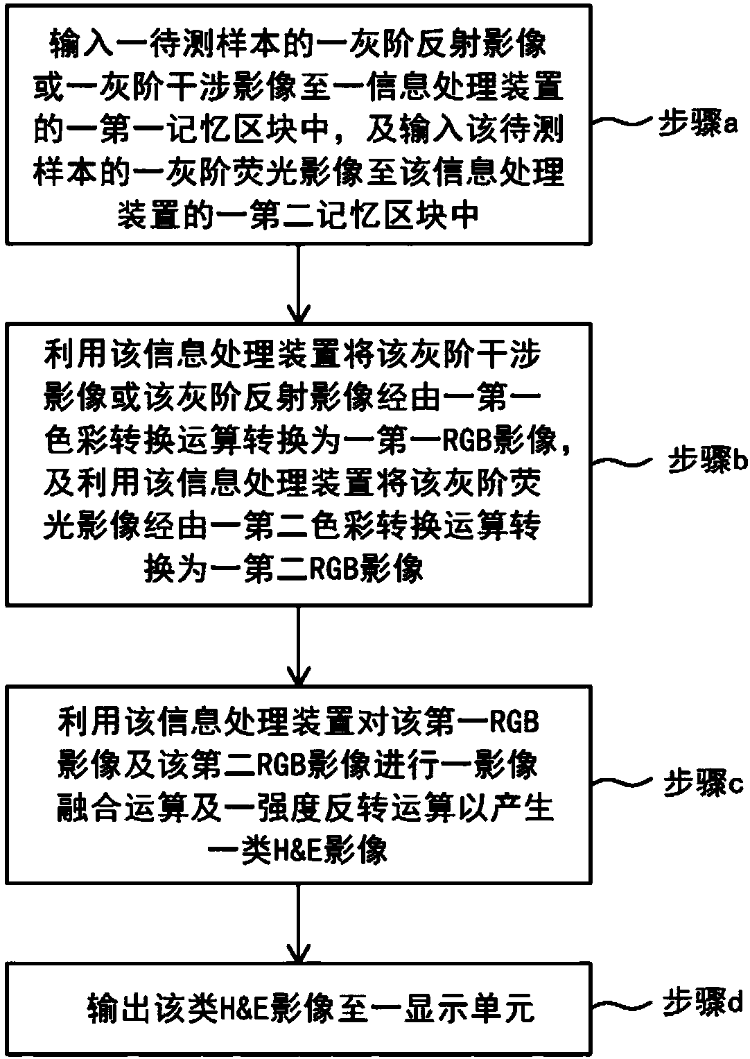
[0037] Please refer to figure 1 , which shows a flowchart of the steps of the method for synthesizing images of biological samples in a preferred embodiment of the present invention.

[0038] As shown in the figure, the image synthesis method of biological samples of the present invention includes the following steps:

[0039] An image synthesis method of a biological sample, comprising the following steps: inputting a gray-scale reflection image or a gray-scale interference image of a biological sample into a first memory block of an information processing device, wherein the gray-scale reflection image Or the gray-scale interference image has a first image resolution, and a gray-scale fluorescence image of the biological sample is input into a second memory block of the information processing device, wherein the gray-scale fluorescence image has a second image resolution, and the resolution of the first image is the same or different from the resolution of the second image ...


PUM


Abstract

Provided in the invention is an image synthesis method for a biological sample. The method comprises steps: inputting a grayscale reflection image or a grayscale interference image of a biological sample into a first memory block of an information processing device and inputting a grayscale fluorescence image of the biological sample into a second memory block of the information processing device;converting the grayscale reflection image or the grayscale interference image into an RGB reflection or interference image by the information processing device based on a first color conversion operation and converting the grayscale fluorescence image into an RGB fluorescence image by the information processing device based on a second color conversion operation; carrying out image fusion operation and intensity inversion operation on the RGB reflection or interference image and the RGB fluorescence image by the information processing device to generate one kind of H&E image; and outputting the H&E image to a display unit. In addition, the invention also discloses an optical system using the image synthesis method for a biological sample.

Description

technical field [0001] The present invention relates to an image synthesis method of an optical system, in particular to an optical slice device using optical interference scanning microscopy or laser scanning confocal microscopy, which is used for image detection of living tissue or confirmation of tissue entry in a biological sample bank. Recognition of tissue cell pattern before bank. Background technique [0002] Living tissue (In Vivo) refers to human tissue that has not been isolated from the body, and is considered to be the most consistent, natural, and original state with the current biological condition of the human body. General patients often need to use tools for imaging inspection to track changes in tissues due to outpatient needs. Among the currently used tools, ultrasound is the tool with a high resolution and can confirm the changes in the internal tissue at a depth of centimeters. It can roughly determine the high and low density of the tissue, and then u...

Claims


Application Information

Patent Timeline
IPC IPC(8): G01N21/01G01N21/64
CPCG01N21/01G01N21/6402G01N21/6486
Inventor 许光裕蔡建中林赐恩
Owner ACUSOLUTIONS INC
Why Eureka
Try Eureka