Simulated Post-Contrast T1-Weighted Magnetic Resonance Imaging

a post-contrast, magnetic resonance imaging technology, applied in the field of system and non-invasive magnetic resonance imaging, can solve the problems of increased patient time, increased risk, and many heart abnormalities that cannot be fully assessed, and achieve the effect of removing common operational and procedural errors

Active Publication Date: 2019-04-25
IMAGING BIOMETRICS
View PDF0 Cites 11 Cited by
  • Summary
  • Abstract
  • Description
  • Claims
  • Application Information

AI Technical Summary

Benefits of technology

[0023]In another embodiment, an image analogous to post-contrast (T1+C) image is generated without the use of an exogenous CA is aut

Problems solved by technology

Also, scans showing the function of blood vessels in real time can be carried out using CAs and many heart abnormalities can only be fully assessed as well.
Although minor, risks do exist.
Proper injection/administration of the CA requires additional patient time (not to mention slight

Method used


Image

  • Simulated Post-Contrast T1-Weighted Magnetic Resonance Imaging
  • Simulated Post-Contrast T1-Weighted Magnetic Resonance Imaging
  • Simulated Post-Contrast T1-Weighted Magnetic Resonance Imaging

Examples


Embodiment Construction

[0029]Selected embodiments of the present invention will now be explained with reference to the figures and flow diagrams. It will be apparent to those skilled in the art from this disclosure that the following descriptions of the embodiments of the present invention are provided for illustration only and not for the purpose of limiting the invention as defined by the appended claims and their equivalents.

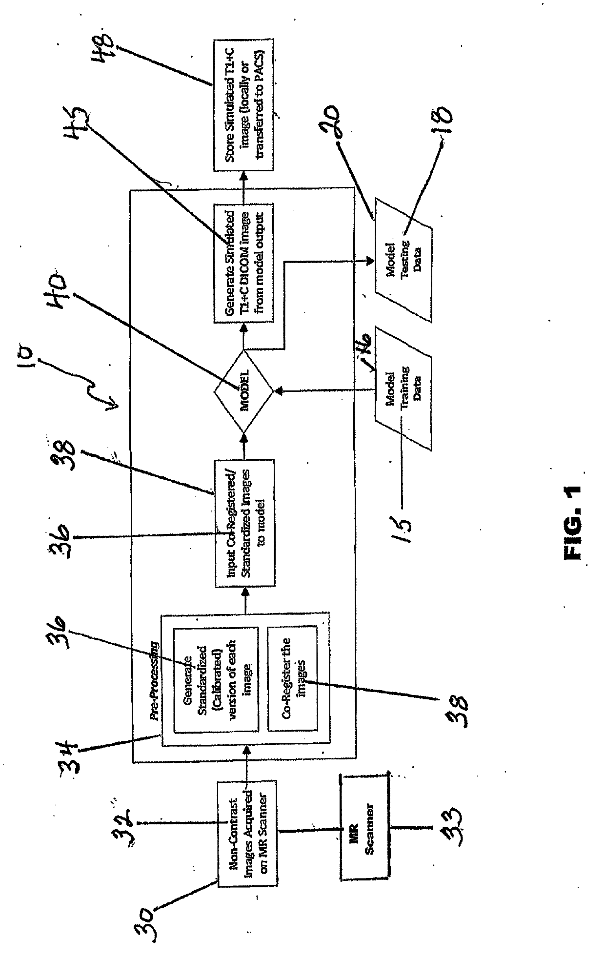
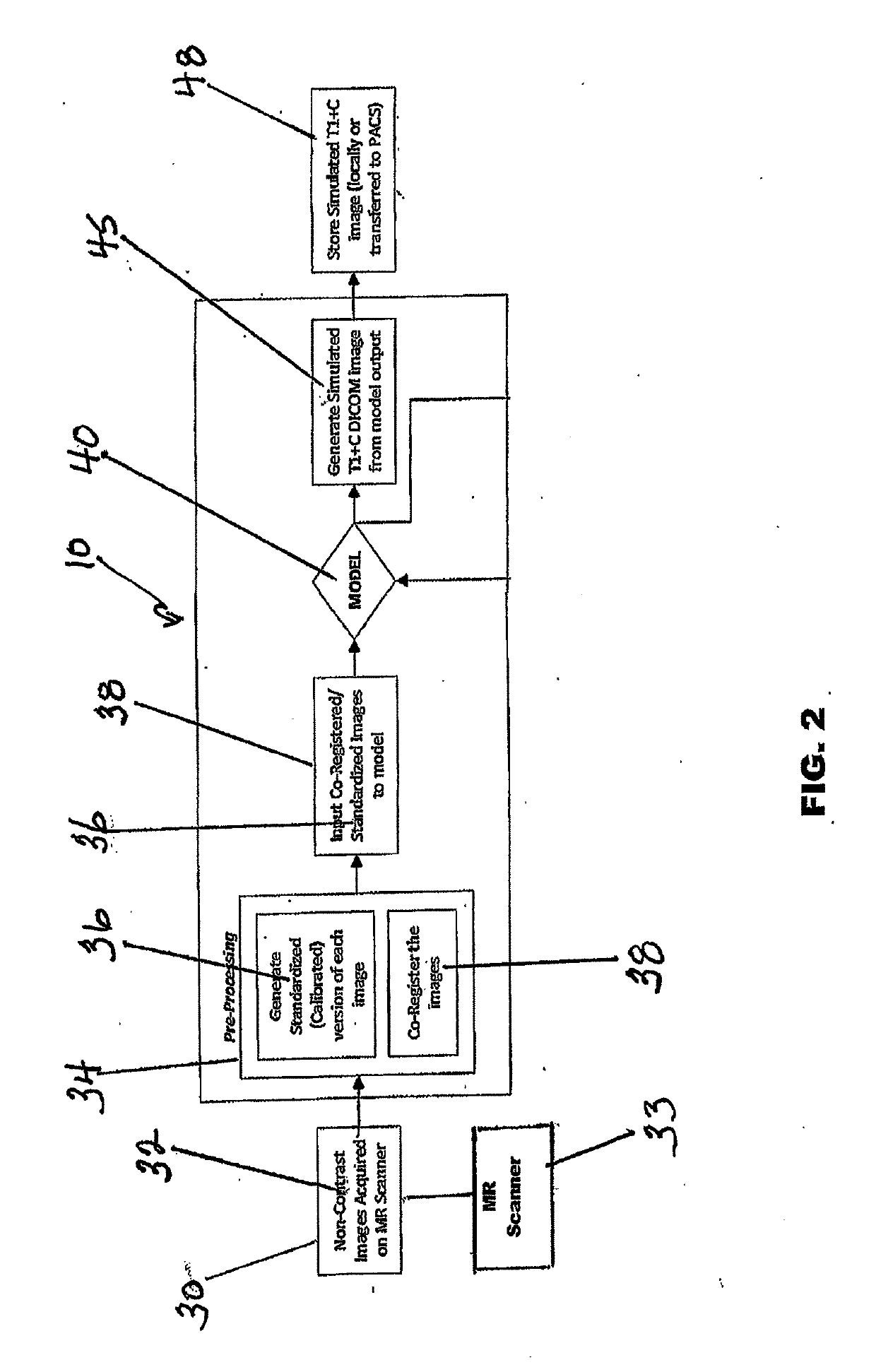
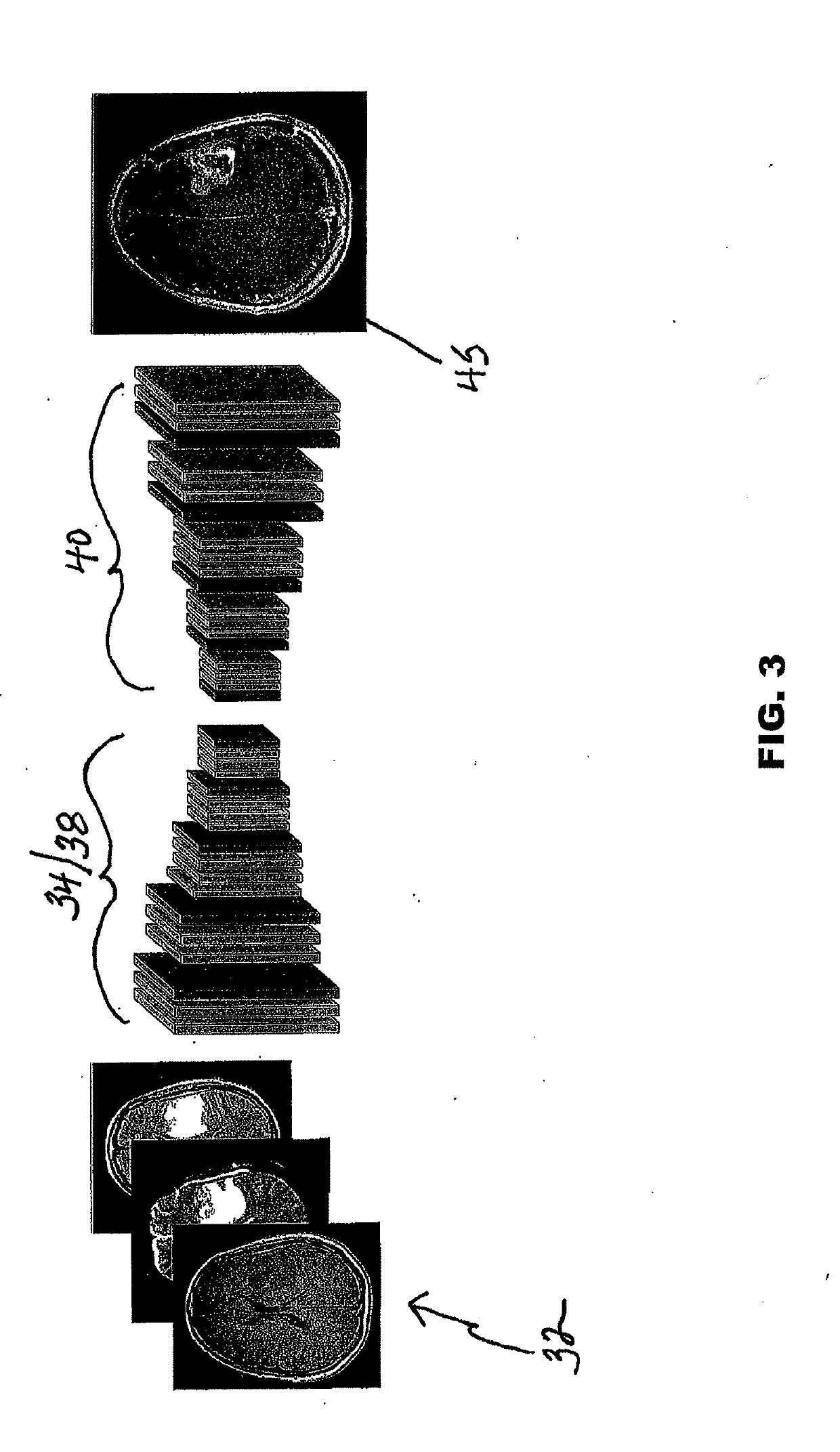
[0030]The visual information in MR images of brain lesions is provided by the change in tissue relaxivity due to the accumulation of contrast agent (CA) material in extravascular tissues caused by a permeable (“leaky”) blood-brain barrier arising from tumor angiogenesis or other disease processes. This information may also be available from a combination of anatomical and functional imaging performed prior to any introduction of exogenous CA. Machine learning is a field of artificial intelligence (AI) which uses statistical methods to impart to computer systems the ability to learn...


PUM


Abstract

A system and method for generating simulated post-contrast T1-weighted magnetic resonance (MR) images without the use of exogenous contrast material based upon patient-specific non-contrast MR images using machine learning/artificial intelligence techniques to train the system to generate post-contrast T1-weighted magnetic resonance images based upon retrospectively collected non-contrast MR images of various sequence types including T1-weighted, T2-weighted, FLAIR (Fluid-Attenuated Inversion Recovery), and/or DWI (Diffusion-Weighted Imaging).

Description

CROSS-REFERENCE TO RELATED APPLICATIONS[0001]This application claims priority to U.S. Provisional Patent Application Ser. No. 62 / 575,048 filed on Oct. 20, 2017.FIELD OF THE INVENTION[0002]The present invention relates generally to magnetic resonant imaging (MRI). More specifically, the present invention related to systems and non-invasive MRI methodologies that provide clearly-defined tissue contrast in MRI images without the use of contrast agents.BACKGROUND OF THE INVENTION[0003]Magnetic resonance imaging (MRI) relies on the relaxation properties of excited hydrogen nuclei in water and lipids to create images. When the target object to be imaged is placed in a uniform magnetic field, the forces in the magnetic field cause the spins of atomic nuclei having a non-zero spin to align in a particular manner with the applied magnetic field. By way of example, hydrogen atoms have a simple spin (½) and therefore align either parallel or anti-parallel to the magnetic field. A radio frequen...

Claims


Application Information

Patent Timeline
IPC IPC(8): G06T5/00
CPCG06T5/009G06T2207/20081G06T2207/30004G06T2207/10088G06T2207/20208G06T7/0016G06T2207/20084G06T2207/30016G16H50/50G16H30/40G06T11/00
Inventor JENSEN, TODD R.
Owner IMAGING BIOMETRICS
Why Eureka
Try Eureka