Non-invasive wearable heart valve monitor

a heart valve and monitor technology, applied in the field of heart valve function monitoring, can solve the problems of heart valve malfunction, heart valve loss, and various problems that can affect the patient's health, and achieve the effect of low-cost and low-power

Active Publication Date: 2019-05-02
EDWARDS LIFESCIENCES CORP
View PDF1 Cites 1 Cited by
  • Summary
  • Abstract
  • Description
  • Claims
  • Application Information

AI Technical Summary

Benefits of technology

The present invention provides a wearable patch for monitoring heart valve function using simple discrete transducers instead of a phased array. The patch is designed to be secured to the patient's chest and can include one or more EKG electrodes, accelerometer, and microphone. The device can calculate blood flow parameters for each heart valve using the ultrasonic signals received from the transducers. The invention also includes a system for monitoring heart valve function by applying a transducer patch to the patient's chest and using a cell phone or remote computer to receive and display the blood flow parameters. The technical effects of the invention include improved accuracy and efficiency in monitoring heart valve function and the ability to do so in a cost-effective and low-power manner.

Problems solved by technology

The operation of the heart, and thus the patient's health, may be seriously impaired if any of the heart valves is not functioning properly.
Various problems can develop with heart valves for a number of clinical reasons.
Heart valves may lose their ability to close properly due to dilation of an annulus around the valve or a flaccid, prolapsed leaflet.
The leaflets may also have shrunk due to disease, such as rheumatic disease, thereby leaving a gap in the valve between the leaflets.
Regurgitation may seriously impair the function of the heart since more blood will have to be pumped through the regurgitating valve to maintain adequate circulation.
Heart valve regurgitation decreases the efficiency of the heart, reduces blood circulation, and adds stress to the heart.
In early stages, heart valve regurgitation leaves a person fatigued and short of breath.
If left unchecked, the problem can lead to congestive heart failure, arrhythmias, or death.
Such phased arrays can provide data that can be processed to create images of the heart and local blood flows, but require trained operator input and assessment and are relatively complex and expensive, and often require relatively high power levels.

Method used


Image

  • Non-invasive wearable heart valve monitor
  • Non-invasive wearable heart valve monitor
  • Non-invasive wearable heart valve monitor

Examples


Embodiment Construction

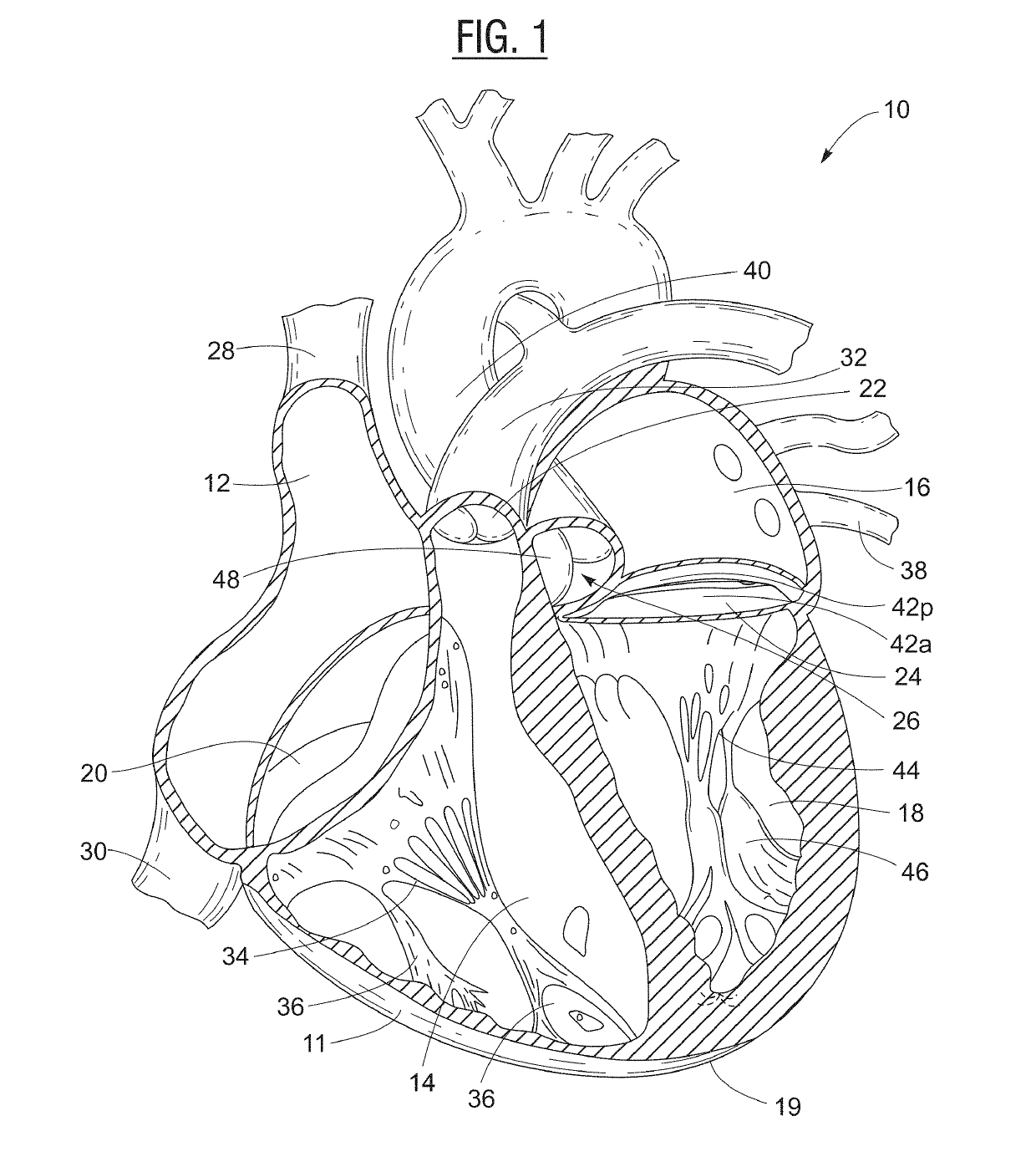
[0033]A cross-sectional view of a human heart 10 is depicted in FIG. 1. The heart 10 has a muscular heart wall 11, an apex 19, and four chambers: right atrium 12; right ventricle 14; left atrium 16; and left ventricle 18. Blood flow is controlled by four main valves: tricuspid valve 20; pulmonary valve 22; mitral valve 24; and aortic valve 26. Blood flows through the superior vena cava 28 and the inferior vena cava 30 into the right atrium 12 of the heart 10. The right atrium 12 pumps blood through the tricuspid valve 20 (in an open configuration) and into the right ventricle 14. The right ventricle 14 then pumps blood out through the pulmonary valve 22 and into the pulmonary artery 32 (which branches into arteries leading to the lungs), with the tricuspid valve 20 closed to prevent blood from flowing from the right ventricle 14 back into the right atrium. Free edges of leaflets of the tricuspid valve 20 are connected via the right ventricular chordae tendinae 34 to the right ventri...


PUM


Abstract

The invention is a device, system, and method for monitoring function of heart valves. The invention includes a disposable patch having simple transducers and simple processing for monitoring heart valve performance via echo Doppler and processing the data to determine blood flow velocities and / or blood flow gradients.

Description

CROSS-REFERENCE TO RELATED APPLICATIONS[0001]This application claims priority to U.S. Provisional Application No. 62 / 579,709, filed Oct. 31, 2018, the contents of which are incorporated herein in their entirety.FIELD OF THE INVENTION[0002]The present invention relates to monitoring heart function, and, more particularly, to methods and apparatuses for the monitoring of heart valve function by positioning a non-invasive monitoring device on the exterior chest wall.BACKGROUND OF THE INVENTION[0003]In vertebrate animals, the heart is a hollow muscular organ having four pumping chambers: the left and right atria and the left and right ventricles, each provided with its own one-way outflow valve. The natural heart valves are identified as the aortic, mitral (or bicuspid), tricuspid and pulmonary valves. The valves separate the chambers of the heart, and are each mounted in an annulus therebetween. The annuluses comprise dense fibrous rings attached either directly or indirectly to the at...

Claims


Application Information

Patent Timeline
IPC IPC(8): A61B8/06A61B8/00A61B5/0245A61B5/0205A61B5/00A61B7/00
CPCA61B8/065A61B8/4281A61B5/0245A61B5/0205A61B5/746A61B5/0022A61B7/003A61B2560/0214A61B2562/0219A61B5/0816A61B2560/0223A61B5/021A61B5/022A61B2505/07A61B8/02A61B8/04A61B8/06A61B8/0883A61B8/4236A61B8/4455A61B8/4472A61B8/4477A61B8/488A61B8/5223A61B8/565A61B8/58A61B8/4494A61B8/0833A61B8/0841A61B8/085A61B8/4427A61B8/4488A61B8/585A61B5/113G16H50/30
Inventor KEIDAR, YARON
Owner EDWARDS LIFESCIENCES CORP
Why Eureka
Try Eureka