

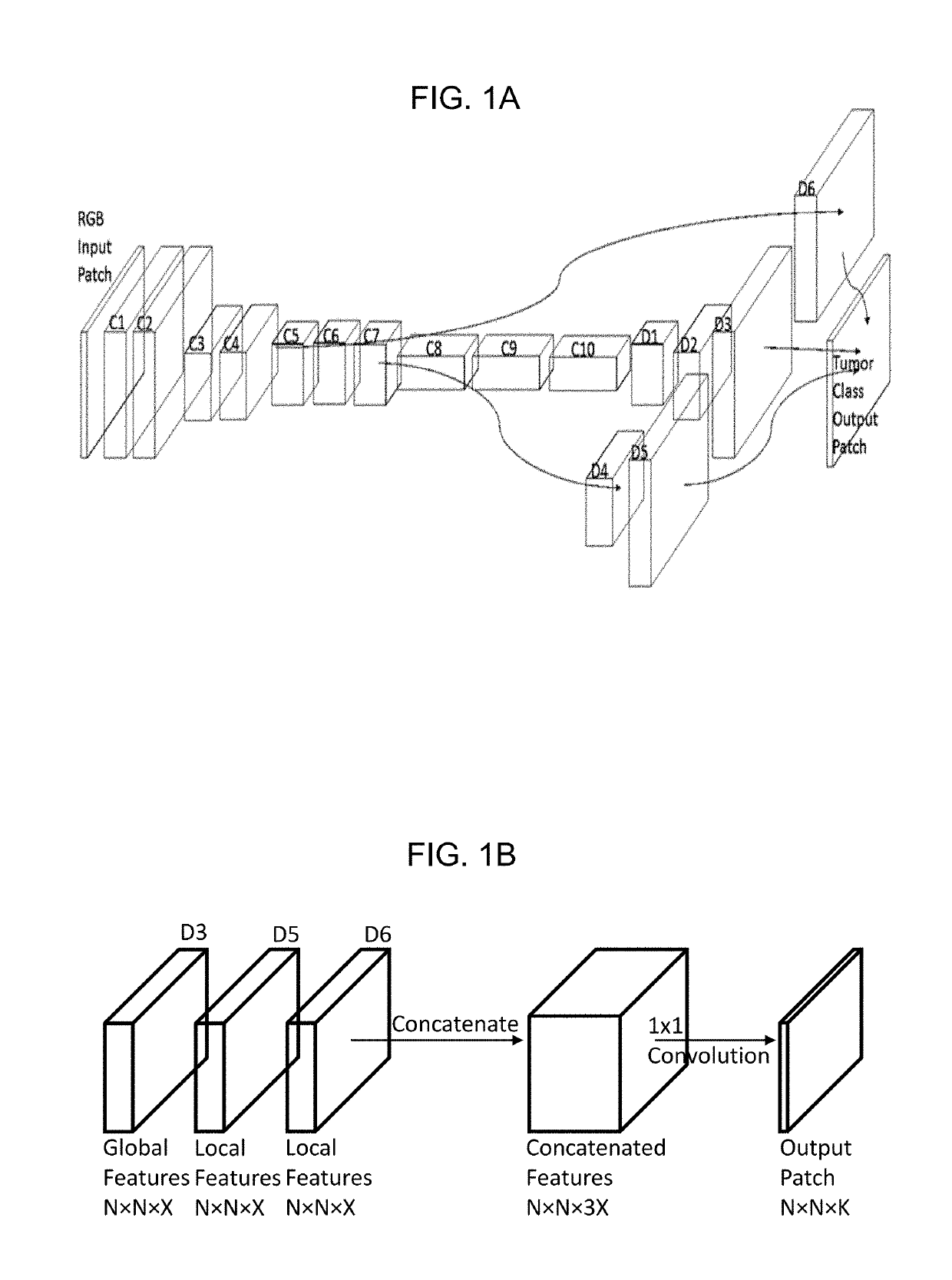
Processing of histology images with a convolutional neural network to identify tumors
a convolutional neural network and tumor technology, applied in image enhancement, medical/anatomical pattern recognition, instruments, etc., can solve the problems of incredibly difficult computer automation to the required level of accuracy and reduced reproducibility
- Summary
- Abstract
- Description
- Claims
- Application Information
AI Technical Summary
Benefits of technology
Problems solved by technology
Method used
Image

Examples
example 1
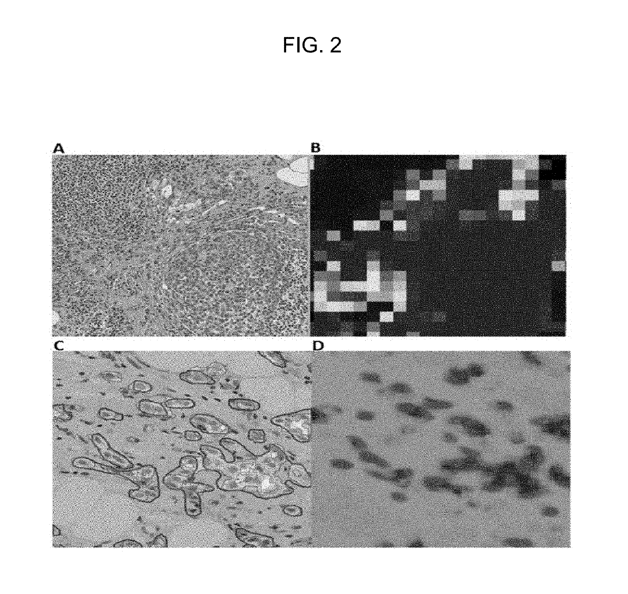
[0094]FIG. 2 is in color and shows the difference between patch-level predictions such as those generated by Google's CNN solution for the Camelyon competition (Liu et al. 2017 [4]) and the pixel-level predictions generated by our CNN.
[0095]Tiles A and B in FIG. 2 are copied from FIG. 7 of Liu et al 2017 [4], whereas Tiles C and D are comparable tiles for an example according to our invention.
[0096]Tile A is a patch from an H&E-stained WSI in which the cluster of larger, dark purple cells in the bottom right quadrant is a tumor, while the smaller dark purple cells are lymphocytes.
[0097]Tile B is a tumor probability heatmap generated by the CNN of Liu et al 2017 [4] which the authors state accurately identifies the tumor cells, while ignoring the connective tissue and lymphocytes.
[0098]Tile C is a raw image patch from an example WSI to which the CNN method embodying the invention is applied. As well as the raw image, Tile C shows outlines drawn manually by a pathologist (solid red pe...
example 2
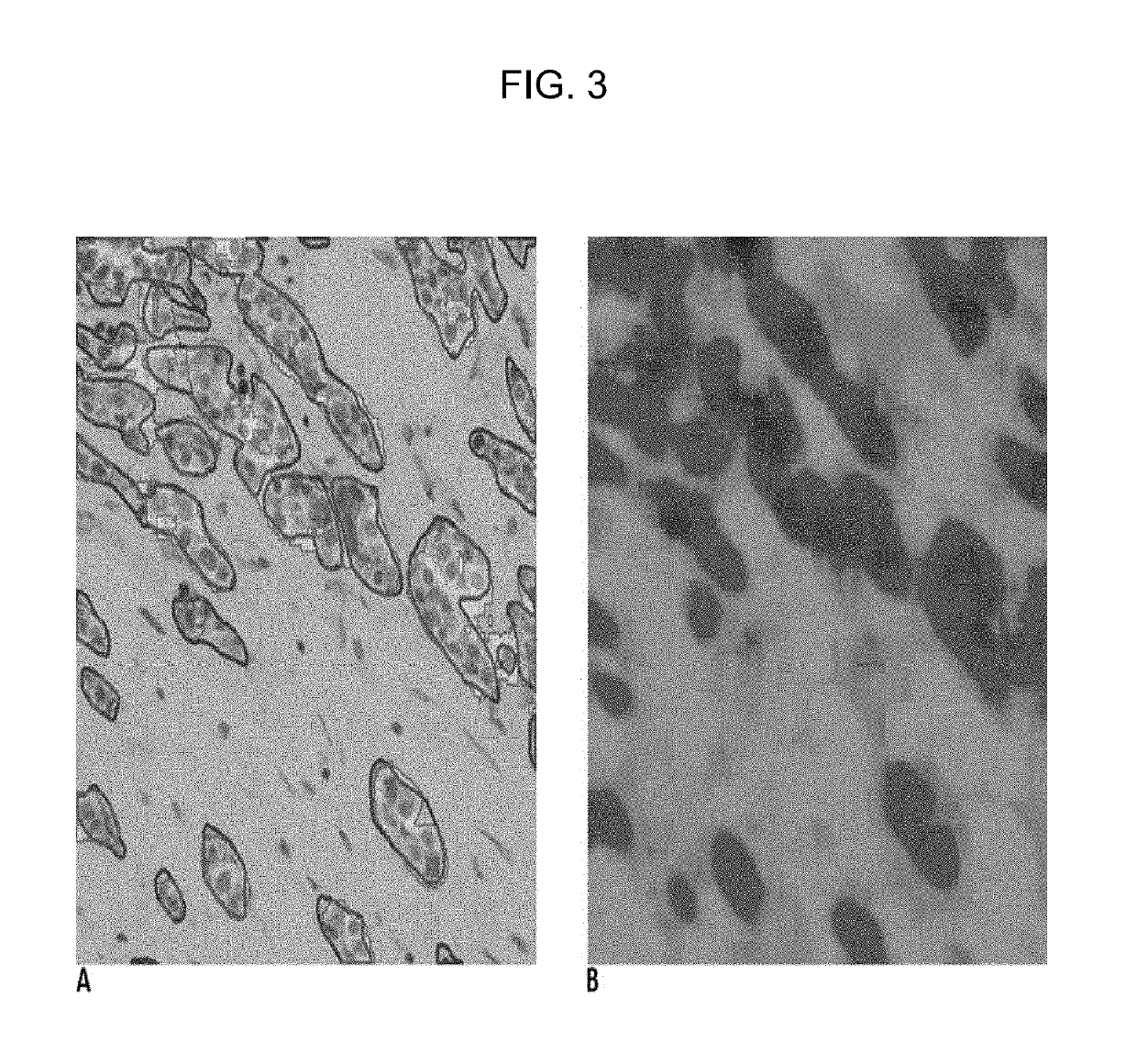
[0100]FIG. 3 is in color and shows an example of the input RGB image patch (Tile A on the left) and the final output tumor probability heat map (Tile B on the right).
[0101]Tile A additionally shows the pathologist's manual outlining of invasive tumors (red outlines) and in addition overlays of our neural network's predictions (shaded pink and yellow areas) as separately shown in Tile B.
[0102]Tile B is a tumor probability heatmap generated by our CNN. For our heatmap, different (arbitrarily chosen) colors indicate different classes, namely green for non-tumor, reddish-brown for invasive tumor (shown pink in Tile A), and blue for in situ tumor (shown yellow in Tile A). Once again, it can be seen how our approach of pixel-level prediction produces areas with smooth perimeter outlines. Moreover, it can be seen how the CNN predictions are compatible with the pathologist's manual marking. In addition, the CNN provides a further distinction between invasive and non-invasive (in situ) tissu...
PUM
 Abstract
Description
Claims
Application Information