
Medical imaging apparatus and method for providing a range of parameters for operation thereof

a technology of medical imaging and parameters, applied in the field of medical imaging apparatus and methods for providing a range of parameters for operation thereof, can solve problems such as invalid quantitative analysis, and achieve the effect of time-efficient and effectiv

Active Publication Date: 2019-08-01
SIEMENS HEALTHCARE GMBH
View PDF3 Cites 0 Cited by
  • Summary
  • Abstract
  • Description
  • Claims
  • Application Information

AI Technical Summary

Benefits of technology

The present invention provides an improved method for analyzing medical image data sets quantitatively in a time-efficient and effective way. The method involves setting a range of potential parameters for operating a medical imaging apparatus to acquire a medical image data set, and using an algorithm to perform a quantitative analysis of the data set based on the patient information. This reduces the likelihood of invalid quantitative analysis resulting from the execution of the algorithm. The method also allows the user to flexibly adapt the parameter range for recording the medical image data set, based on the algorithm and the patient's status. This avoids recording a medical image data set that cannot be appropriately analyzed and saves time for subsequent analysis. The method can be used with magnetic resonance tomography scans and can be adapted to the cardiac cycle for increasing the probability of recording a medical image data set with proper T1-values.

Problems solved by technology

For example, the quantitative analysis is considered to be invalid when a bias exceeds a specific value.

Method used


Image

  • Medical imaging apparatus and method for providing a range of parameters for operation thereof
  • Medical imaging apparatus and method for providing a range of parameters for operation thereof

Examples


Embodiment Construction

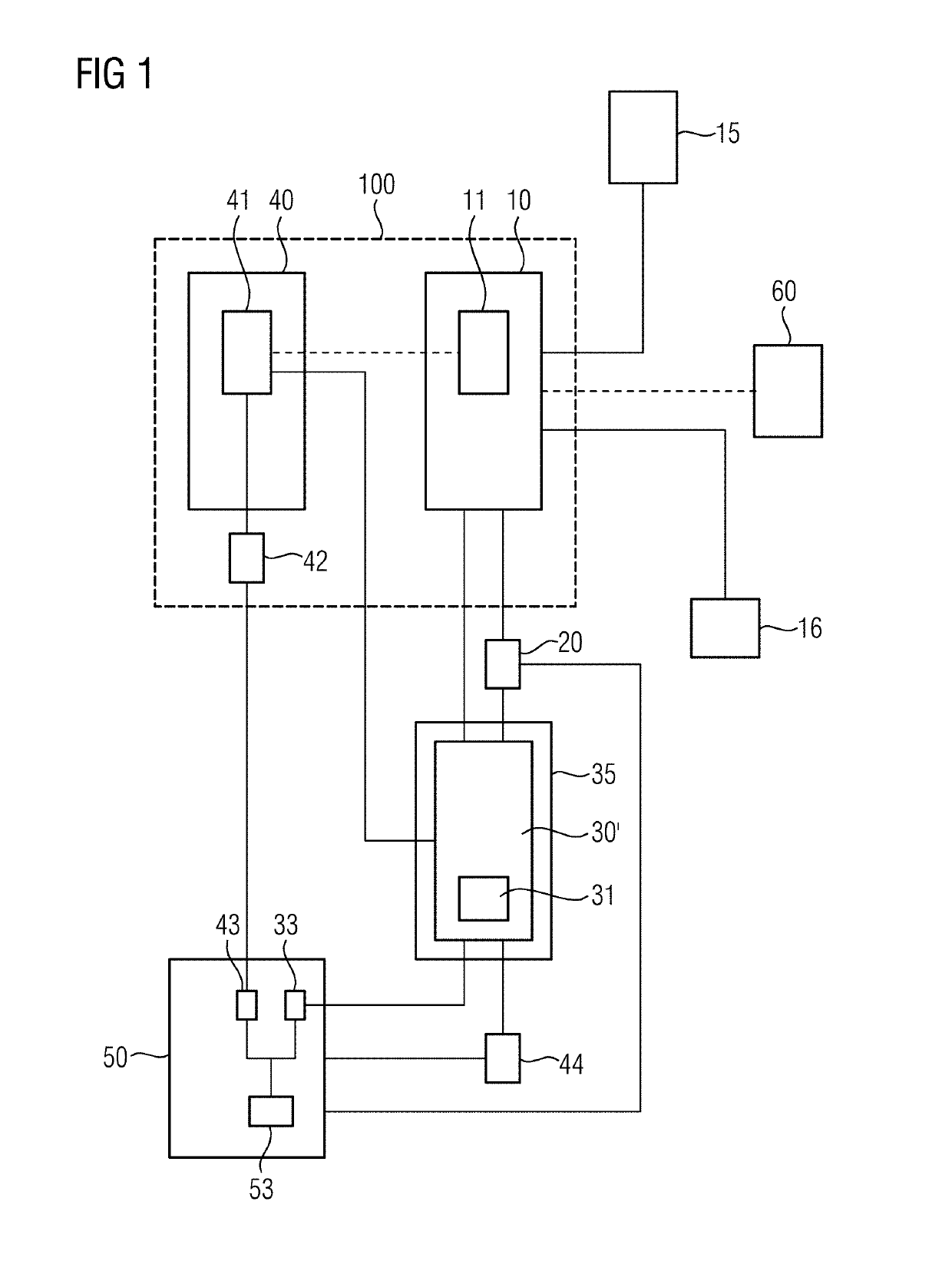
[0023]In FIG. 1, a block diagram is presented that schematically illustrates the method for providing a range of potential parameters 11 that can be used for recording a future medical image data set 31 according to a preferred embodiment of the present invention. For instance, such a medical image data set 31 is provided by recording 30 a magnetic resonance tomography (MRT) scan by means of a medical imaging device 35. Besides visualizing the medical image data sets 31 to show the shape or inner structure of an organ, in particular of parts of the organ, it is preferably provided that further specifications of the medical image data set 31 are specified by key values 42. Preferably, these key values 42 are mapped or assigned to specific regions of the visualized medical image data set and for example help to differentiate different kinds of tissues. Thereby it is thinkable that a visualization 33 of the medical image data set 31 and a further visualization 43 representing the key v...


PUM


Abstract

In a method and medical imaging apparatus for providing a range of potential parameters that can be used for recording a future medical image data set, an algorithm is provided to a computer for performing a quantitative analysis of the future medical image data set. The computer is also provided with patient information specifying a status of a patient. In order to reduce a probability of an invalid quantitative analysis, the range of potential parameters is set in the computer by performing the quantitative analysis depending on the patient information.

Description

BACKGROUND OF THE INVENTIONField of the Invention[0001]The present invention concerns a method for providing a range of potential parameters that can be used for recording a medical image data set, as well as a corresponding medical imaging apparatus, and a non-transitory, computer-readable data storage medium.Description of the Prior Art[0002]Recording medical image data sets, for example by operating a magnetic resonance tomography scanner, is well known. Typically, the recorded medical image data sets are reconstructed for visualization thereof on a screen in order to observe an inner structure of an organ or a part of an organ. In some cases, it is even possible to further specify sub-parts of the visualized parts of an organ, preferably for evaluating a particular tissue. For this purpose, besides the reconstruction, a quantitative analysis of the medical image data set is performed. Such a quantitative analysis preferably results in a set of key values, each of which further s...

Claims


Application Information

Patent Timeline
IPC IPC(8): G16H30/40G16H30/20
CPCG16H30/40G16H30/20G06T7/0012G16H40/63A61B6/481A61B5/055
Inventor GREISER, ANDREAS
Owner SIEMENS HEALTHCARE GMBH
Learn More