Novel Anti-pd-1 antibodies
a technology of anti-pd1 antibodies and antibodies, which is applied in the field of new anti-pd1 antibodies, can solve the problems that the existing therapies may not be all satisfactory, and achieve the effects of reducing tumor volume, good tolerability, and high in vivo anti-tumor activity
- Summary
- Abstract
- Description
- Claims
- Application Information
AI Technical Summary
Benefits of technology
Problems solved by technology
Method used
Image
Examples
example 1
Hybridoma Generation
[0247]1.1 Immunogen generation: DNAs encoding PD-1 and PD-L1 ECD or full length were synthesized and inserted into the expression vector pcDNA3.3. Max-prep the plasmid DNAs and the inserted DNA sequences were verified by sequencing. Fusion proteins PD-1 ECD and PD-L1 ECD containing various tags, including human Fc, mouse Fc and His tags, were obtained by transfection of human PD-1 ECD gene into CHO-S or HEK293 cells. After 5 days, supernatants harvested from the culture of transiently transfected cells were used for protein purification. The fusion proteins were purified and quantitated for usage of immunization and screening.
[0248]1.2 Stable cell lines establishment. In order to obtain tools for antibody screening and validation, we generated PD-1 and PD-L1 transfectant cell lines. Briefly, CHO-K1, 293F or Ba / F3 cells were transfected with pCND3.3 expression vector containing full-length PD-1 or PD-L1 using Lipofectamine 2000 Transfection kit according to manufa...
example 2
Hybridoma Cell Sequence and Fully Human Antibody Characterization
[0255]2.1 Antibody hybridoma cell sequence: RNAs were isolated from monoclonal hybridoma cells with Trizol reagent. The VH and VL of PD-1 antibodies were amplified as following protocol: briefly, RNA is first reverse transcribed into cDNA using a reverse transcriptase as described here, Reaction system (20 μl):
10× RT Buffer2.0 μl25× dNTP Mix (100 mM)0.8 μl10× RT Random Primers / oligodT / specific primer2.0 μlMultiScribe ™ Reverse Transcriptase1.0 μlRNase Inhibitor1.0 μlRNA 2 μgNuclease-free H2O to 20.0 μl
[0256]Reaction Condition
Step1Step2Step3Step4Temperature2537854Time10 min120 min5 min∞
[0257]The resulting cDNA is used as templates for subsequent PCR amplification using primers specific for interested genes. The PCR reaction was done as following procedure;
cDNA 1 μlEx PCR buffer 5 μldNTP 2 μlExTaq0.5 μlP1(25pM)0.5 μlP2(25pM)0.5 μlddH2O40.5 μl
[0258]Reaction Condition:
[0259]94° C. 3 min
94°C.30s60°C.30s}30cycles[0260]7...
example 3
an Antibody Characterization
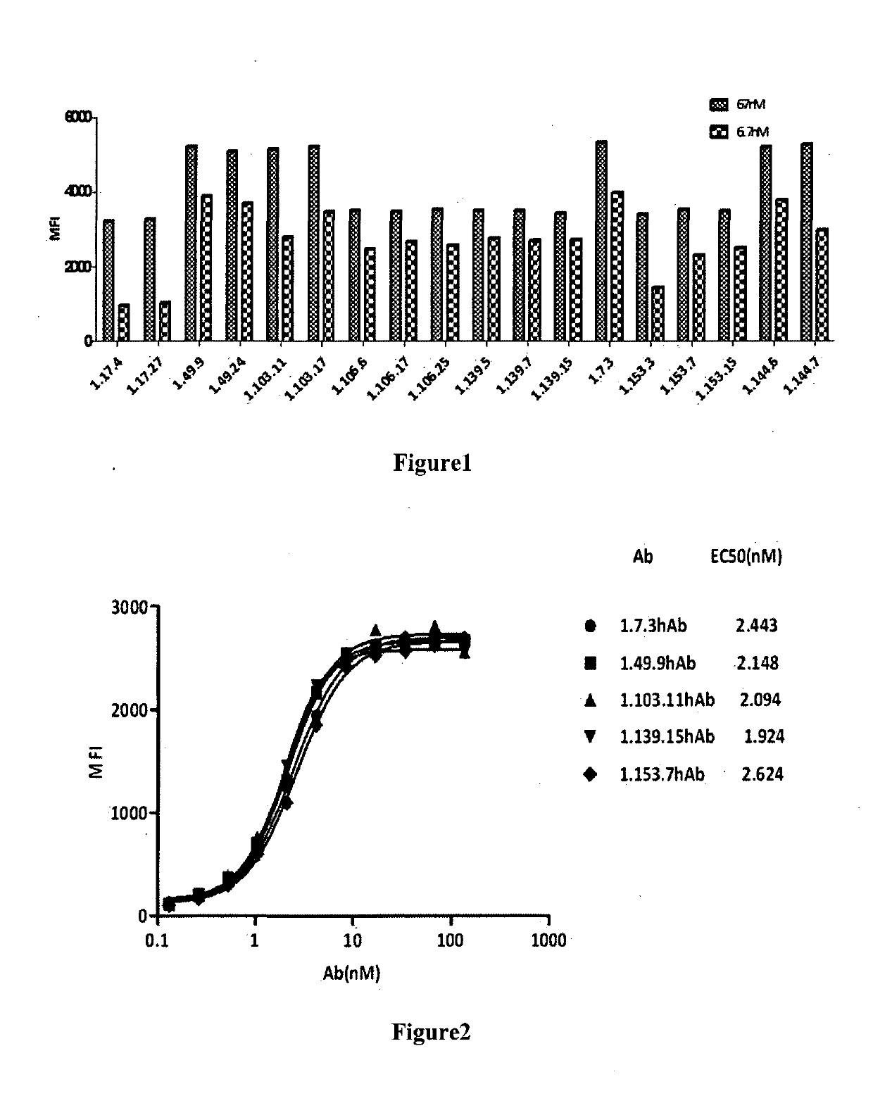
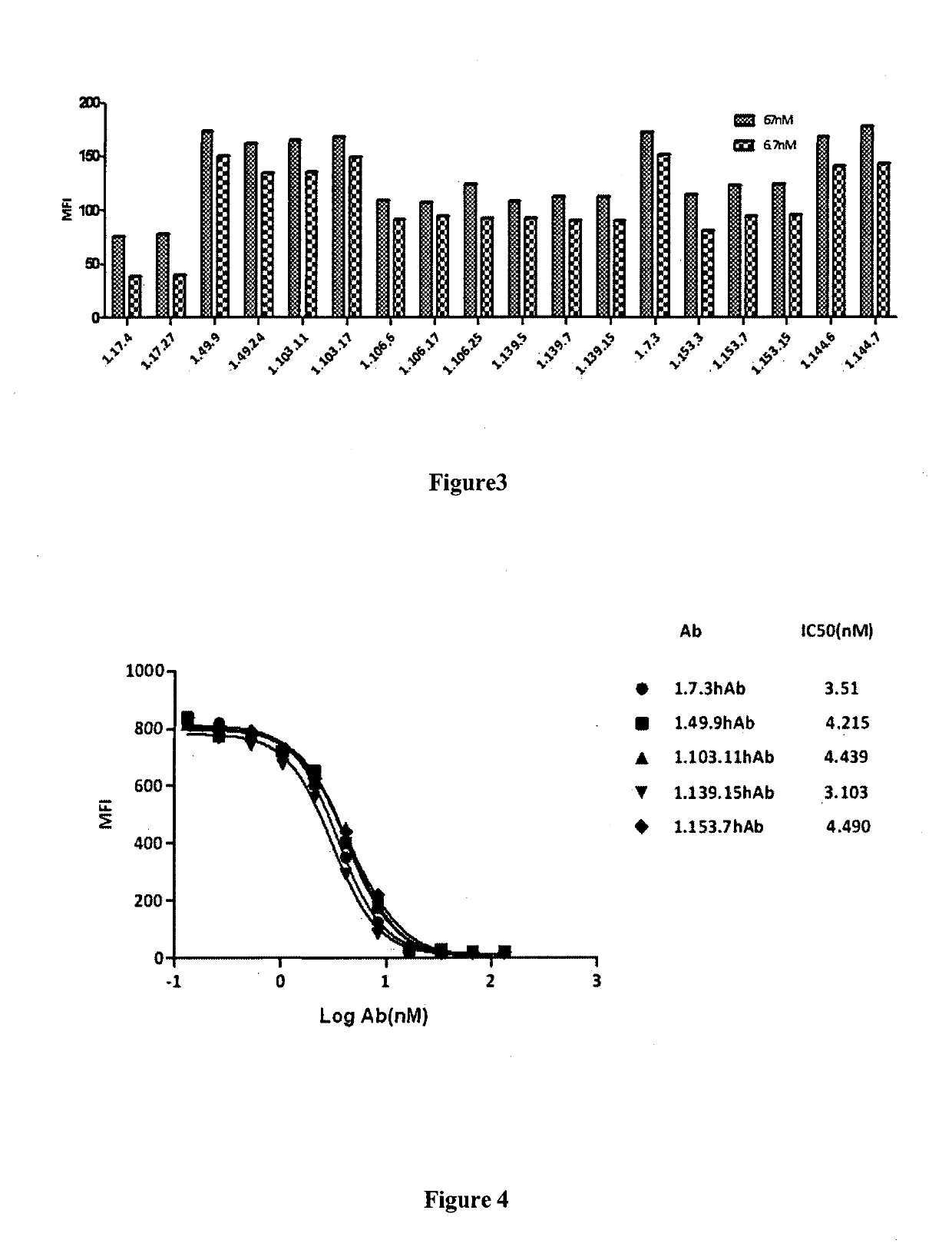
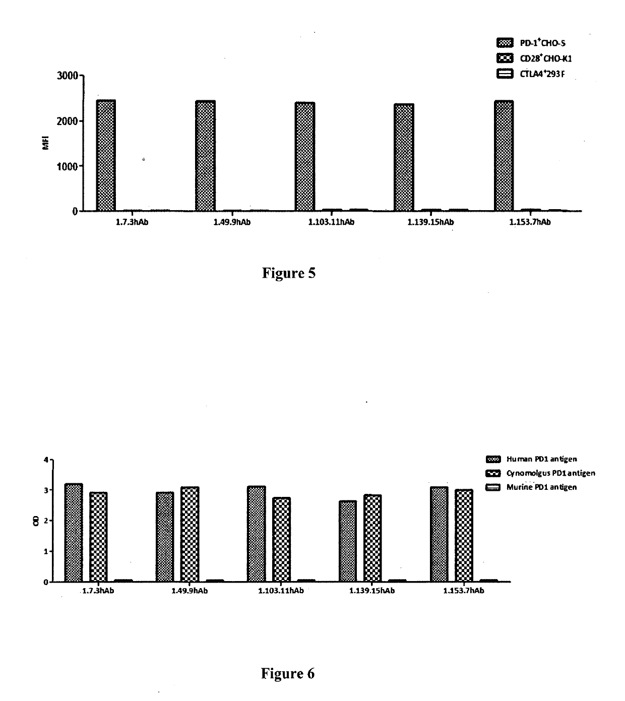
[0265]3.1 Binding affinity of PD-1 antibodies to cell surface PD-1 molecules tested by flow cytometry (FACS): Antibody binding affinity to cell surface PD-1 was performed by FACS analysis. CHO-S cells expressing human PD-1 were transferred in to 96-well U-bottom plates (BD) at a density of 5×10⁵ cells / ml. Tested antibodies were 1:2 serially diluted in wash buffer (1×PBS / 1% BSA) and incubated with cells at 4° C. for 1 h. The secondary antibody goat anti-human IgG Fc FITC (3.0 moles FITC per mole IgG, (Jackson Immunoresearch Lab) was added and incubated at 4° C. in the dark for 1 h. The cells were then washed once and resuspended in 1×PBS / 1% BSA, and analyzed by flow cytometery (BD). Fluorescence intensity will be converted to bound molecules / cell based on the quantitative beads Quantum™ MESF Kits, Bangs Laboratories, Inc.). KD was calculated using Graphpad Prism5. FIG. 2 shows the binding of the fully human PD-1 antibodies (i.e. 1.7.3 hAb, 1.49.9 hAb, 1.10...
PUM
| Property | Measurement | Unit |
|---|---|---|
| Mass | aaaaa | aaaaa |
| Mass | aaaaa | aaaaa |
| Fraction | aaaaa | aaaaa |
Abstract
Description
Claims
Application Information
 - R&D
- Intellectual Property
- Life Sciences
- Materials
- Tech Scout
- Unparalleled Data Quality
- Higher Quality Content
- 60% Fewer Hallucinations
Browse by: Latest US Patents, China's latest patents, Technical Efficacy Thesaurus, Application Domain, Technology Topic, Popular Technical Reports.
© 2025 PatSnap. All rights reserved.Legal|Privacy policy|Modern Slavery Act Transparency Statement|Sitemap|About US| Contact US: help@patsnap.com


