

Materials and methods for treating regional pain
a technology of regional pain and materials, applied in the field of regional pain treatment materials and methods, can solve the problems of affecting life quality and no cure for regional pain, and achieve the effect of inducing neurolysis of the drg
- Summary
- Abstract
- Description
- Claims
- Application Information
AI Technical Summary
Benefits of technology
Problems solved by technology
Method used
Image

Examples
example 1
d DRG-Targeted Delivery of Resiniferatoxin (RTX)
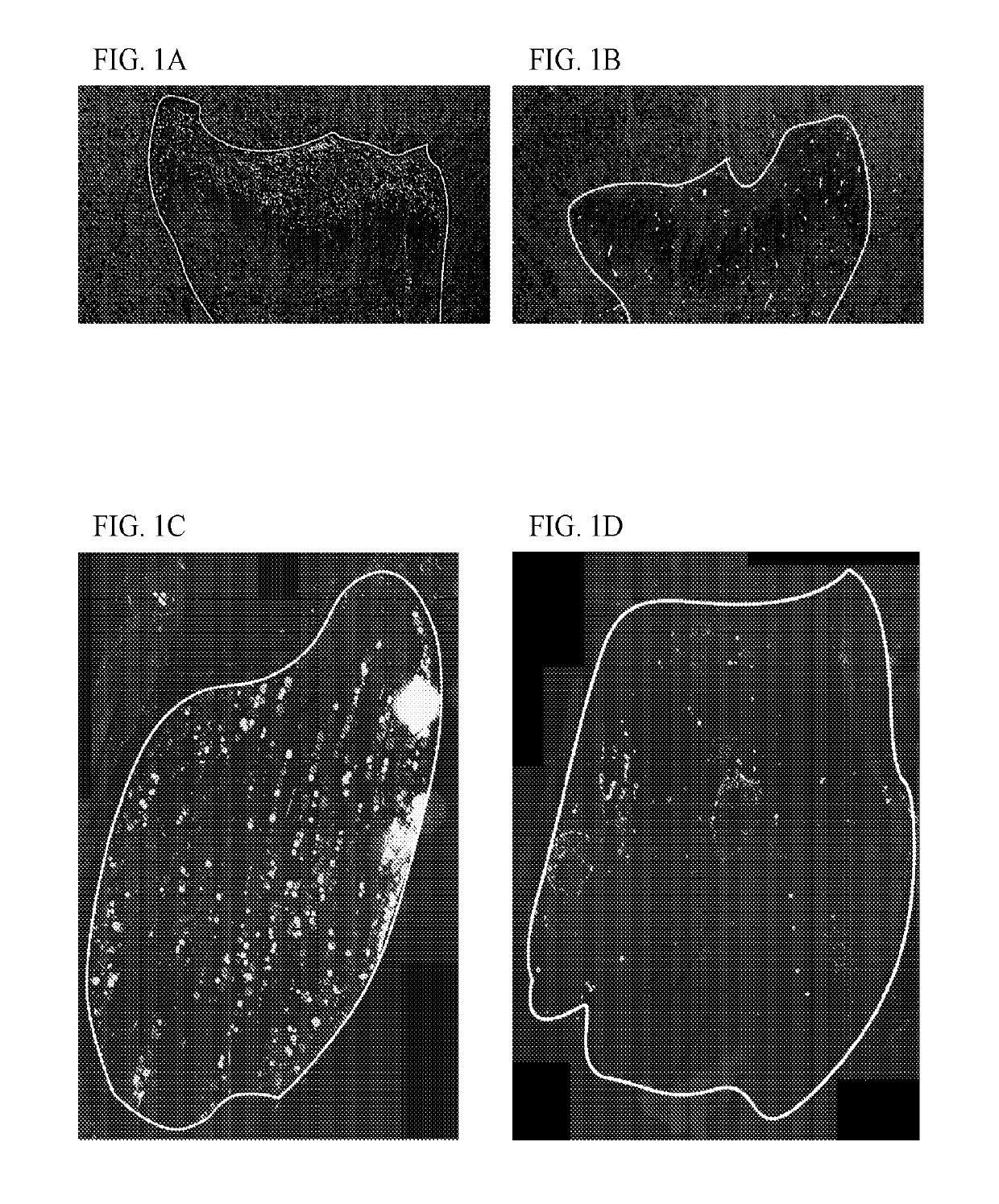
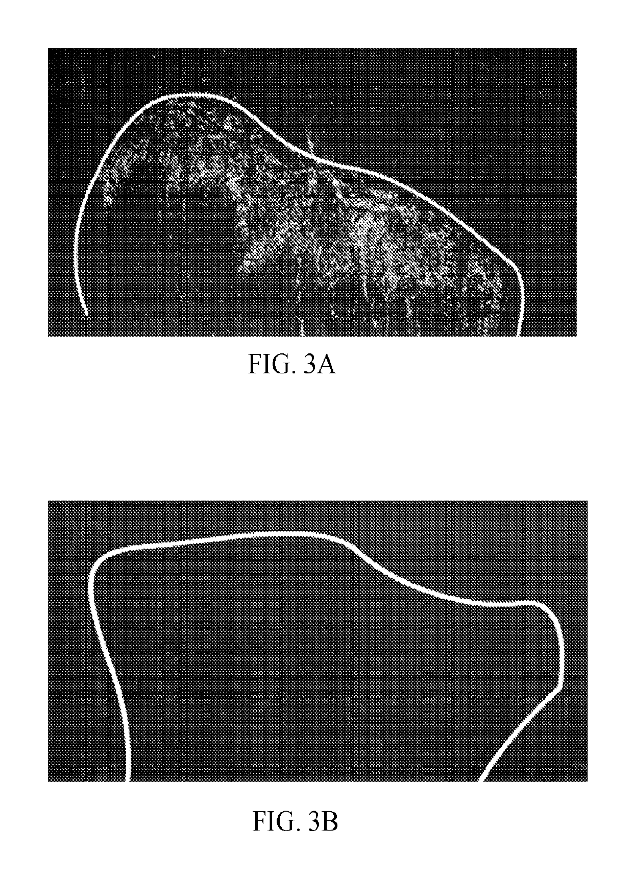
[0039]Domestic swine were chosen because the porcine lumbar spinal column resembles the human lumbosacral anatomy. Swine were sedated with Telazol and Xylazine, intubated and kept under deep isoflurane inhalation anesthesia for the duration of the procedure titrated to effect. Swine were placed in an MRI scanner (GE 3T, GE Healthcare). Overview MRI imaging was obtained of the lumbosacral spine. DRG at the L4, L5, S1, and S2 level were outlined (FIG. 1A).
[0040]The guide needle was passed through the skin lateral to the midline and incrementally advanced ventromedially toward the DRG intraprocedural MRI imaging monitored advancement of the needle, and any deviations from the optimal trajectory were corrected. When the needle tip was visualized directly adjacent to the dorsal aspect of the DRG, the stylet of the guide needle was withdrawn. The stepped stylet was then inserted through the guide needle. The length of the stepped stylet exce...
example 2
d DRG-Targeted Delivery of Resiniferatoxin (RTX)
[0042]An incision was made in the overlying skin at an anatomical level affected by pain in a patient. The subcutaneous fat and musculature were incised and reflected, exposing the bony lamina. Using appropriate instruments, a portion of the lamina was removed to expose the neural elements. The meningeal sleeve surrounding the DRG and spinal nerve was surgically exposed by microdissection of bone and tissue, which may require extending the laminectomy dissection, using standard techniques such as blunt or sharp dissection with meticulous hemostasis. Thereby the DRG was exposed and grossly visualized. A convection enhanced delivery needle was placed into the exposed DRG. RTXcap was injected in a preparation that also contains a contrast agent (e.g., in this case DAPI) allowing direct visualization of analgesic agent injection by eye sight with or without a surgical loupe or surgical operating microscope.
[0043]Convection enhanced deliver...
example 3
d DRG-Targeted Delivery of a Nucleic Acid Molecule
[0047]The subject is placed in an MRI scanner. Overview MRI imaging is obtained of the lumbosacral spine. The guide needle is passed through the skin lateral to the midline and incrementally advanced ventromedially toward the DRG. Intraprocedural MRI imaging monitors advancement of the needle, and any deviations from the optimal trajectory is corrected. When the needle tip is visualized directly adjacent to the dorsal aspect of the DRG, the stylet of the guide needle is withdrawn. The stepped stylet is then inserted through the guide needle. The length of the stepped stylet exceeds the length of the guide needle and therefore only the stepped tip of the stylet but not the Quincke tip of the guide needle penetrates the DRG parenchyma. The stepped stylet is then withdrawn and replaced by the stepped needle. The prior insertion of the stepped stylet prevents clogging of the narrow needle tip. A formulation consisting of phosphate buffer...
PUM
| Property | Measurement | Unit |
|---|---|---|
| Volume | aaaaa | aaaaa |
| Volume | aaaaa | aaaaa |
| Volume | aaaaa | aaaaa |
Abstract
Description
Claims
Application Information