

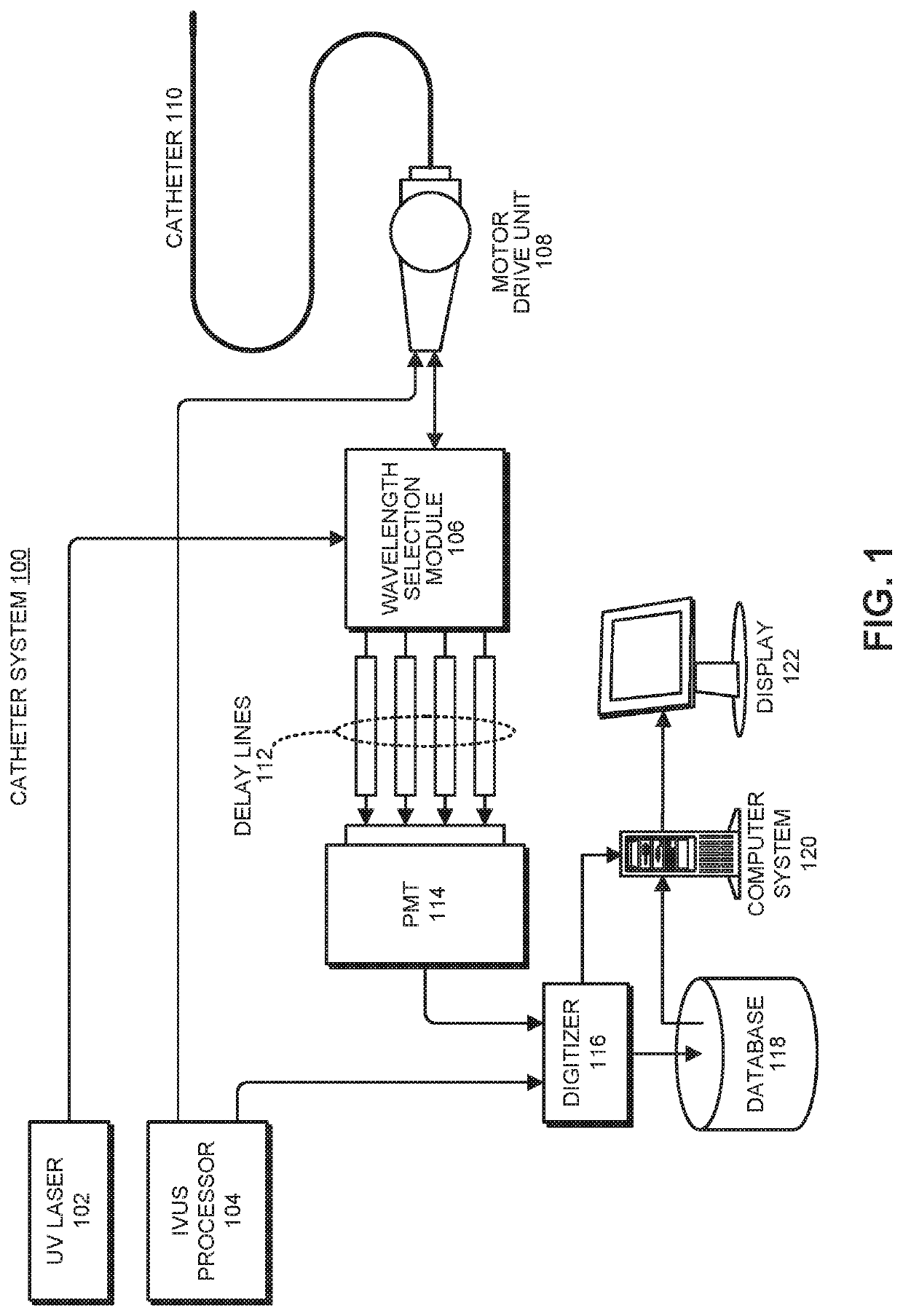
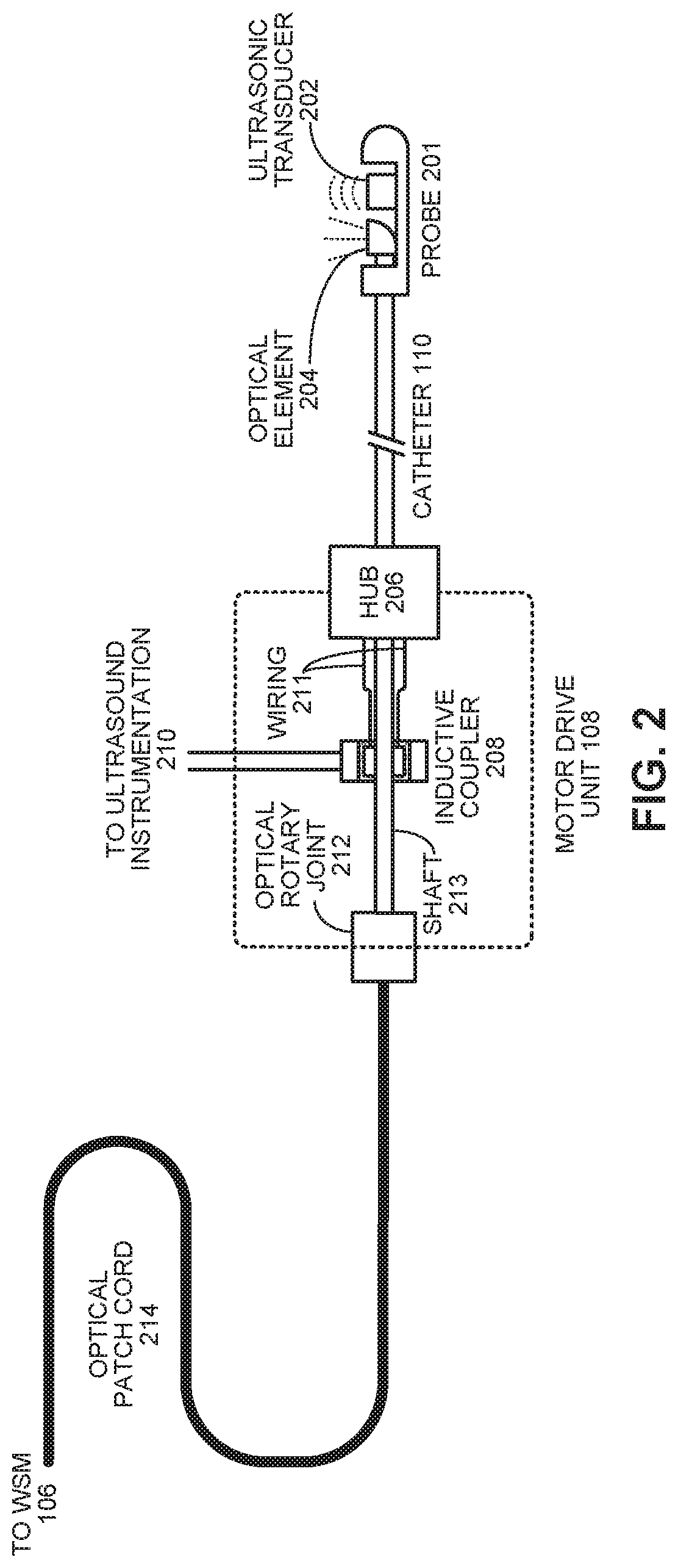
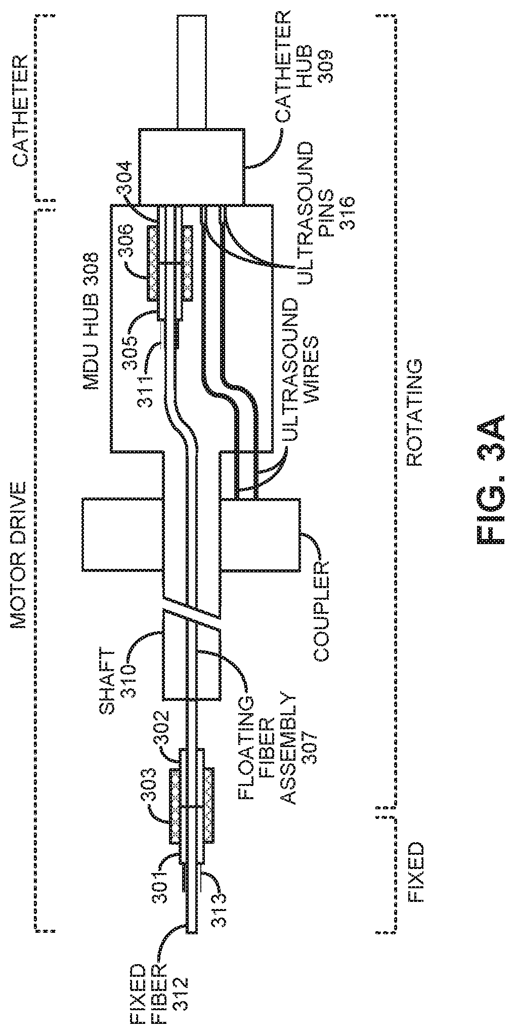
Single catheter system that provides both intravascular ultrasound and fluorescence lifetime imaging
a single catheter and lifetime imaging technology, applied in the field of catheter-based vascular imaging systems, can solve the problems of limited imaging penetration depth, inability to reliably determine the size of deep plaque cores or outward remodeling, and inability to detect rupture-prone thin-cap fibroatheromas (tcfas),
- Summary
- Abstract
- Description
- Claims
- Application Information
AI Technical Summary
Benefits of technology
Problems solved by technology
Method used
Image

Examples
Embodiment Construction
[0023]The following description is presented to enable any person skilled in the art to make and use the present embodiments, and is provided in the context of a particular application and its requirements. Various modifications to the disclosed embodiments will be readily apparent to those skilled in the art, and the general principles defined herein may be applied to other embodiments and applications without departing from the spirit and scope of the present embodiments. Thus, the present embodiments are not limited to the embodiments shown, but are to be accorded the widest scope consistent with the principles and features disclosed herein.
[0024]The data structures and code described in this detailed description are typically stored on a computer-readable storage medium, which may be any device or medium that can store code and / or data for use by a computer system. The computer-readable storage medium includes, but is not limited to, volatile memory, non-volatile memory, magneti...
PUM
 Abstract
Description
Claims
Application Information