
Monoclonal antibody inhibiting immunosuppressive functions of pathogens, antigen-binding fragment thereof, and hybridomas producing such antibody

a technology of monoclonal antibodies and pathogens, which is applied in the direction of antibacterial agents, fused cells, peptides, etc., can solve the problems chronic infections and the development of gastric pathologies in certain patients, and achieve the suppression of host immunity, block the immunosuppressive functions of pathogens, and prevent the effect of affecting the growth of i

Active Publication Date: 2020-01-09
SAGABIO CO LTD
View PDF0 Cites 0 Cited by
  • Summary
  • Abstract
  • Description
  • Claims
  • Application Information

AI Technical Summary

Benefits of technology

[0008]According to this invention, certain pathogens are capable of suppressing the immunity of their hosts. Such immunosuppressive functions are especially provided by immunosuppressive substances secreted or produced by the pathogens. It is possible to block the immunosuppressive functions of the pathogens by shutting down functions of the immunosuppressive substances. Since the immunosuppressive functions have been shut down, the immunity activities of the host is not suppressed. The unsuppressed or increased immunity functions of the host are then capable of reducing or even eliminating the pathogens.
[0016]The monoclonal antibody of the invention expresses significant inhibitive effects against immunosuppressive activities of particular pathogens and is useful in blocking an immunosuppressive phenomenon caused by the pathogens. In certain embodiments of the present invention, the immunosuppressive function of pathogens is provided by an immunosuppressive substance secreted or produced by the pathogens. In such embodiments, the monoclonal antibody or its antigen-binding fragment of the present invention primarily eliminate the pathogens by inhibiting the immunosuppressive functions of the pathogens, and therefore activating immune responses of the host of the pathogens.

Problems solved by technology

Although a predominant Th1-polarized mucosal immune response is activated in the host, the immune response is not sufficient to mount protective immunity against H. pylori, resulted in chronic infections and development of gastric pathologies in certain patient.
In addition, the administration of an anti-HSP60 antibody was found to interfere with the growth of H. pylori.

Method used


Image

  • Monoclonal antibody inhibiting immunosuppressive functions of pathogens, antigen-binding fragment thereof, and hybridomas producing such antibody
  • Monoclonal antibody inhibiting immunosuppressive functions of pathogens, antigen-binding fragment thereof, and hybridomas producing such antibody
  • Monoclonal antibody inhibiting immunosuppressive functions of pathogens, antigen-binding fragment thereof, and hybridomas producing such antibody

Examples


embodiment 1

and Isolation of PBMC and T-Cells

[0069]Human peripheral blood mononuclear cells (PBMCs) from healthy donors were isolated by density gradient centrifugation using Ficoll-Paque Plus (GE Healthcare, Uppsala, Sweden) and resuspended in RPMI-1640 with 10% inactivated fetal calf serum and 1% penicillin-streptomycin. For monocyte depletion, PBMCs were cultured in 10-cm dishes at a density of 106 / ml overnight for monocyte attachment. The suspended cells were then collected by centrifugation at 1500 rpm for 15 min. Total T-cells were isolated from PBMCs by negative selection using a magnetic sorting device (Miltenyi Biotec, MA, USA). Briefly, PBMCs were incubated with a cocktail of biotin-conjugated antibodies, followed by microbead-conjugated anti-biotin Abs for magnetic depletion. T-cells were eluted according to the manufacturer's protocols.

embodiment 2

f HpHSP60 on PBMC Proliferation

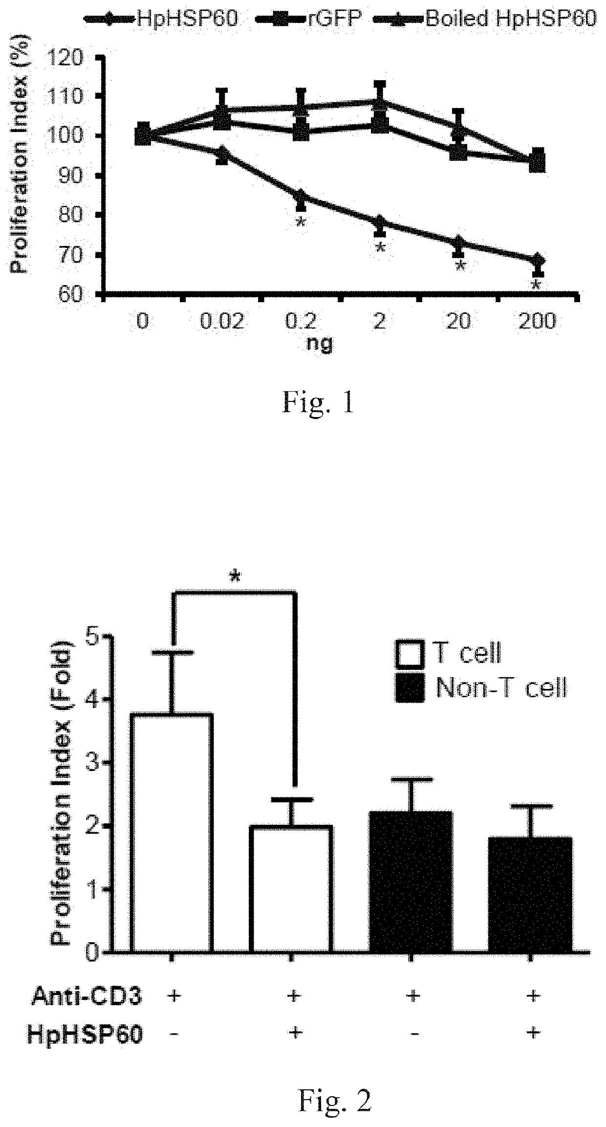
[0070]Proliferation of anti-CD3 mAb-stimulated PBMCs treated with HpHSP60, rGFP or boiled HpHSP60 at different doses was monitored by a cell proliferation assay. To measure cell proliferation, 0.2 ml of cells at 1×10⁶ cells / ml were seeded in each well of an anti-CD3 mAb-precoated 96-well microplate. Cell proliferation was determined by an MTT assay after 96 hours. Results are shown in FIG. 1: experimental results on effects of HpHSP60 on PBMC proliferation. Data shown therein are reported as the proliferation index.

[0071]The cell proliferation index was calculated as follows: Proliferation index (100%)=(OD595 of the anti-CD3+HpHSP60-treated cells) / (OD595 of the anti-CD3-treated cells)*100%. The results that differ significantly from the untreated group are indicated by * (p<0.05) (n=15).

[0072]In FIG. 1, (♦) shows proliferation of T-cells is inhibited, after HpHSP60 is added into the PBMC. (▪) shows rGFP, being a control protein in this experimental sys...

embodiment 3

HpHSP60 on T-Cell Proliferation in PBMC

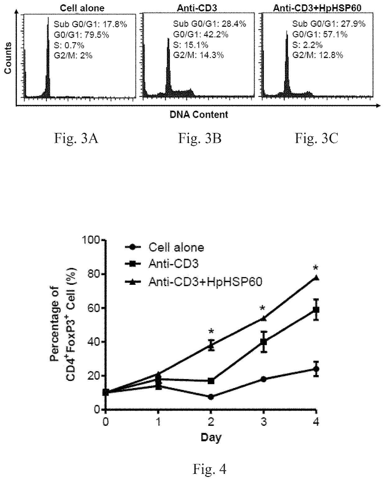
[0073]After treatment with anti-CD3 mAb, PBMCs were treated with or without HpHSP60 (200 ng). T-cells or non-T-cells in PBMCs were identified by CD3 surface marker staining. Cell number was then calculated following a flow cytometer analysis.

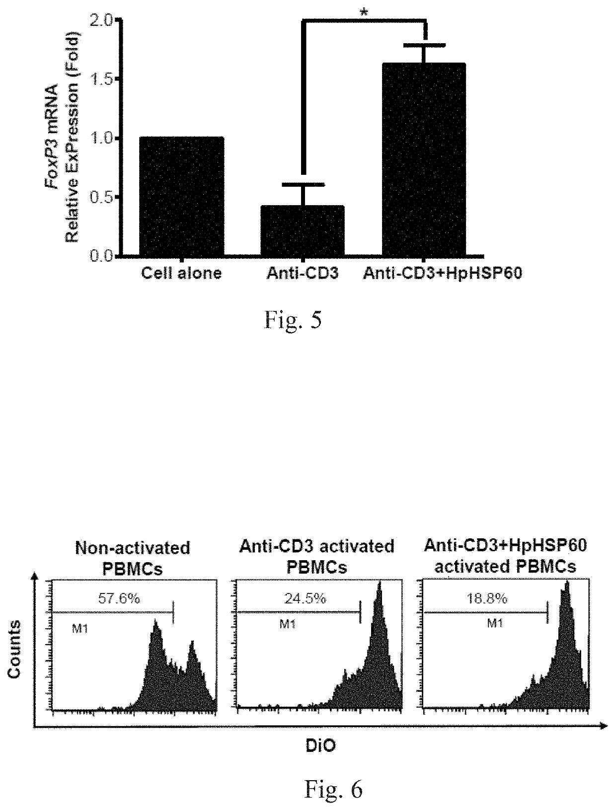
[0074]For CD3 surface marker staining, cells were harvested and stained with 1 μg mouse anti-human CD3 IgG mAbs (OKT3), followed by 0.5 μg rabbit anti-mouse IgG-FITC secondary Abs (Biolegend, CA, USA). For FoxP3 intracellular staining, the cells were harvested and stained with mouse anti-human CD4-FITC mAbs (Biolegend, CA, USA) prior to fixing and permeabilization, followed by intracellular staining with mouse anti-human FoxP3-PE mAbs (BD Biosciences, MA, USA) according to the manufacturer's protocol. For the cell cycle assay, cells were harvested after 72 hours and 106 cells were fixed with 70% ice-cold ethanol. DNA was stained with DNA staining buffer (5% Triton-X 100, 0.1 mg / ml RNase A, and 4 μg / ml propi...


PUM

PropertyMeasurementUnit
temperatureaaaaaaaaaa
heat shockaaaaaaaaaa

Abstract

The present invention relates to a monoclonal antibody that inhibits immunosuppressive functions of pathogens, antigen-binding fragment thereof, and hybridomas producing such antibody. The monoclonal antibody or antigen-binding fragment thereof bind to a peptide consisting an amino acid sequence represented by MEKVGKDGVITVE (SEQ ID NO: 1). The present invention also discloses use of the invented monoclonal antibody or antigen-binding fragment thereof, and method of preparation for such hybridomas.

Description

CROSS REFERENCE TO RELATED APPLICATIONS[0001]This application is a Continuation of co-pending application Ser. No. 15 / 165,723, filed on May 26, 2016, for which priority is claimed under 35 U.S.C. § 120; the entire contents of all of which are hereby incorporated by reference.FIELD OF THE INVENTION[0002]The present invention relates to a monoclonal antibody that inhibits the immunosuppressive functions of a pathogen, antigen-binding fragment thereof, and hybridomas producing such antibody, and especially to a monoclonal antibody that inhibits the immunosuppressive functions provided by an immunosuppressive substance secreted or produced by the pathogen, to enhance the immune system of host of the pathogen, and its antigen-binding fragment and hybridomas producing the antibody.BACKGROUND OF THE INVENTION[0003]Helicobacter pylori (H. pylori) is a Gram-negative bacterium that infects half of the adult population worldwide. The chronic inflammation triggered by H. pylori can lead to vari...

Claims


Application Information

Patent Timeline
Patent Type & Authority Applications(United States)
IPC IPC(8): C07K16/12
CPCC07K2317/565C07K2317/76C07K2317/14C07K16/121A61K2039/505C07K2317/56C07K2317/34A61P31/04C07K16/18C12N5/163A61K39/395
Inventor LIAO, KUANG-WENLIN, YU-LINGJIAN, TING-YAN
Owner SAGABIO CO LTD
Learn More