Artificial neural network and system for identifying lesion in retinal fundus image

a technology of artificial neural network and retinal fundus, applied in image enhancement, instruments, applications, etc., can solve the problem that the method is not ready for clinical applications, and achieve the effect of improving the screening accuracy

Active Publication Date:
View PDF0 Cites 32 Cited by
  • Summary
  • Abstract
  • Description
  • Claims
  • Application Information

AI Technical Summary

Benefits of technology

[0026]According to the present disclosure, disclosed are an artificial neural network and an artificial neural system for identifying a lesion in a retinal fundus image with improved screening accuracy, and a method for identifying a lesion in a retinal fundus image.

Problems solved by technology

However, in the prior art as described above, although deep learning methods have been employed to automatically identify various lesions of diabetic retinopathy using retinal fundus images, such methods are not ready for clinical applications.

Method used


Image

  • Artificial neural network and system for identifying lesion in retinal fundus image
  • Artificial neural network and system for identifying lesion in retinal fundus image
  • Artificial neural network and system for identifying lesion in retinal fundus image

Examples


first embodiment

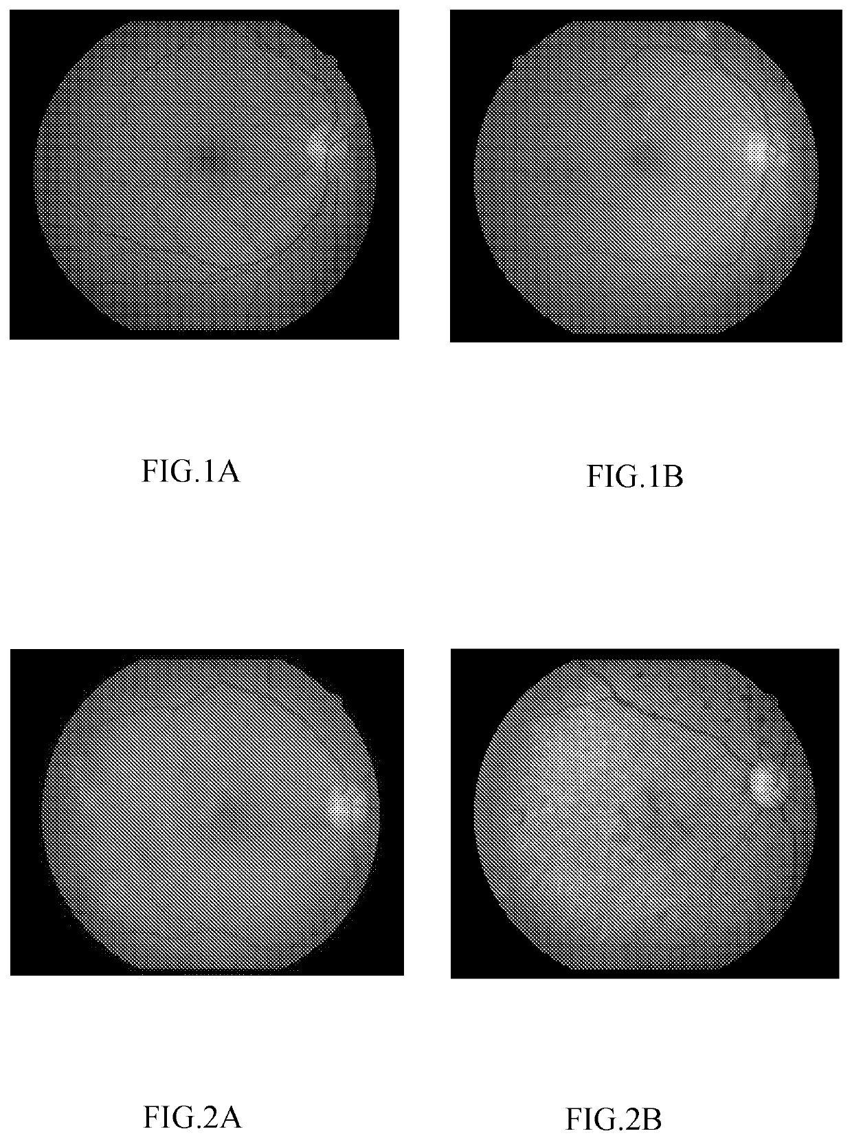
[0044]FIG. 1 is a schematic diagram illustrating lesions in retinal fundus images according to a first embodiment of the present disclosure, wherein FIG. 1(a) shows an exemplary image of normal retinal fundus, and FIG. 1(b) shows an exemplary image of abnormal retinal fundus. FIG. 2 is a schematic diagram illustrating retinal fundus images with retinal fundus lesions according to the first embodiment of the present disclosure, wherein FIG. 2(a) shows an exemplary image of retinal fundus with diabetic retinopathy, and FIG. 2(b) shows an exemplary image of retinal fundus with hypertensive retinopathy.

[0045]In this embodiment, an artificial neural network and an artificial neural network system according to this embodiment systematically study lesion-free retinal fundus images (see FIG. 1(a)) and retinal fundus images with lesions (see FIG. 1(b)), and become capable of determining whether a lesion is present in a retinal fundus image. In addition, in this embodiment, the artificial neu...

second embodiment

[0128]FIG. 10 is a block diagram showing an artificial neural network 10B according to the second embodiment of the present disclosure. FIG. 11 is a diagram illustrating a third neural network 14 according to the second embodiment of the present disclosure. FIG. 12 is a block diagram showing a pre-processing module 31 of the artificial neural network 10B according to the second embodiment of the present disclosure.

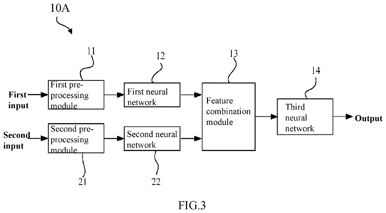
[0129]The difference between the artificial neural network 10B according to this embodiment and the artificial neural network 10A according to the first embodiment is that the artificial neural network 10B comprises a pre-processing module 31, and the third neural network 14 may generate a diagnosis result according to the feature combination set and patient information (see FIG. 10). The artificial neural network 10B according to this embodiment can also improve the accuracy (including sensitivity and specificity) of screening of retinal fundus lesions.

[0130]The feature c...


PUM


Abstract

The present disclosure provides an artificial neural network system for identifying a lesion in a retinal fundus image that comprises a pre-processing module configured to separately pre-process a target retinal fundus image and a reference retinal fundus image taken from a same person; a first neural network (12) configured to generate a first advanced feature set from the target retinal fundus image; a second neural network (22) configured to generate a second advanced feature set from the reference retinal fundus image; a feature combination module (13) configured to combine the first advanced feature set and the second advanced feature set to form a feature combination set; and a third neural network (14) configured to generate, according to the feature combination set, a diagnosis result. By using a target retinal fundus image and a reference retinal fundus image as independent input information, the artificial neural network may simulate a doctor, determining lesions on the target retinal fundus image using other retinal fundus images from the same person as a reference, thereby enhancing the diagnosis accuracy.

Description

FIELD OF THE INVENTION[0001]The present disclosure relates to the field of artificial neural networks, and more particularly to an artificial neural network and an artificial neural network system for identifying a lesion in a retinal fundus image.BACKGROUND OF THE INVENTION[0002]An artificial neural network is a machine learning model that simulates the human brain structure. With the development of artificial neural networks, especially artificial intelligence technology such as deep learning, the application of artificial neural networks in the field of medical imaging diagnosis is drawing more and more attention. By using such an artificial neural network, potential lesions can be automatically determined based on medical images, i.e., automatic screening can be done on the medical images. For example, artificial neural networks such as deep learning have been studied in various fields, such as breast cancer pathological analysis, lung cancer detection, cardiovascular imaging an...

Claims


Application Information

Patent Timeline
Patent Type & Authority Applications(United States)
IPC IPC(8): A61B3/00G06T7/00G06N3/04G06N3/08G06N20/20G06N20/10
CPCG06T2207/30041G06N20/20G06T2207/30096G06N3/0454G06T2207/20081G06N20/10G06T2207/20084A61B3/0025G06T7/0012G06N3/08A61B3/12A61B3/14G06T7/0014G06N3/045
Inventor WANG, JUANXSA, BINBAI, YUJINGLI, XIAOXINHU, ZHIGANGZHAO, YU
Why Eureka
Try Eureka