Medical Image Pre-Processing at the Scanner for Facilitating Joint Interpretation by Radiologists and Artificial Intelligence Algorithms

a technology of medical image data and pre-processing algorithms, which is applied in the field of pre-processing medical image data at the medical image scanner, can solve the problems of large amount of raw medical image data not being used for training ai algorithms, and none of these pre-processing algorithms are informed on the downstream workflow for the resulting medical imag

Active Publication Date: 2020-04-02
SIEMENS HEALTHCARE GMBH
View PDF15 Cites 3 Cited by
  • Summary
  • Abstract
  • Description
  • Claims
  • Application Information

AI Technical Summary

Benefits of technology

[0011]In an embodiment, the second set of pre-processing algorithms includes one or more pre-processing algorithms corresponding to each of a plurality of available downstream automated image analysis algorithms predicted to be applied to the medical image scan, and generating one or more medical images for automated image analysis by applying the second set of pre-processing algorithms to the raw medical image data comprises: generating a respective medical image suited for each of the plurality available downstream automated image analysis algorithms predicted to be applied to the medical image scan using the corresponding one or more pre-processing algorithms in the second set of pre-processing algorithms.
[0012]In an embodiment, the trained machine learning based model is trained based on a training database of training images acquired using a variety of pre-processing algorithms by comparing output interpretations of the training images determined using one or more available downstream automated image processing algorithms with ground truth interpretations of the training images.
[0013]In an embodiment of the present invention, an apparatus for medical image acquisition and pre-processing comprises: means for acquiring raw medical image data by performing a medical image scan of a patient using a medical image scanner; means for acquiring input data associated with the medical image scan of the patient and available downstream automated im

Problems solved by technology

As a result, a large amount of information in the raw medical image data is not being utilized for training the AI algorithm

Method used


Image

  • Medical Image Pre-Processing at the Scanner for Facilitating Joint Interpretation by Radiologists and Artificial Intelligence Algorithms
  • Medical Image Pre-Processing at the Scanner for Facilitating Joint Interpretation by Radiologists and Artificial Intelligence Algorithms
  • Medical Image Pre-Processing at the Scanner for Facilitating Joint Interpretation by Radiologists and Artificial Intelligence Algorithms

Examples


Embodiment Construction

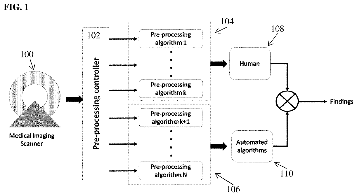
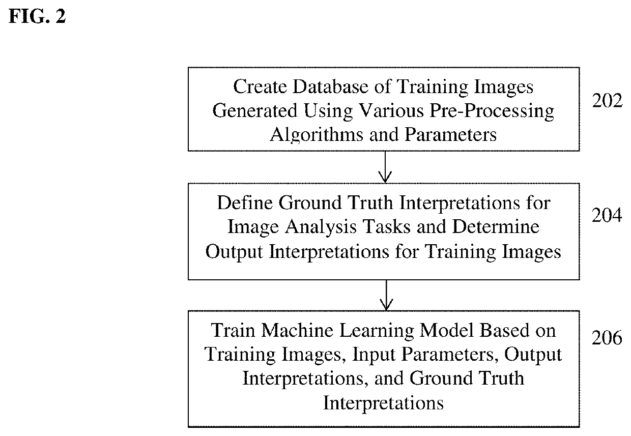
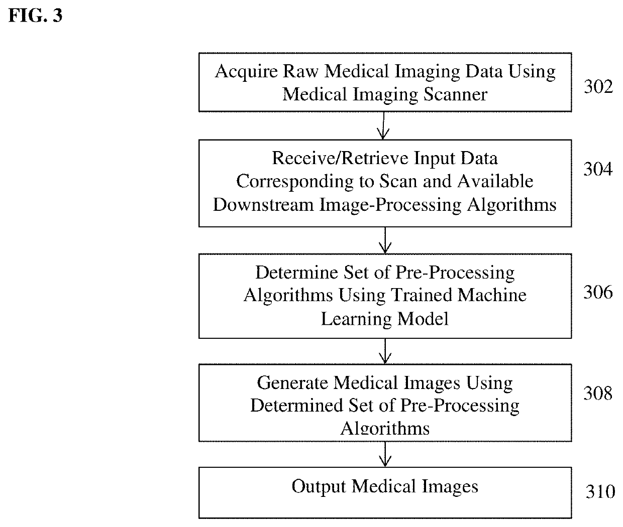
[0020]The present invention relates to medical image pre-processing at the medical image scanner to facilitate joint interpretation of the medical images by radiologists and artificial intelligence algorithms. Embodiments of the present invention provide a method and system for computer-based medical image pre-processing that is automatically tailored based on the downstream tasks to be performed on the acquired medical image data. A digital image is often composed of digital representations of one or more objects (or shapes). The digital representation of an object is often described herein in terms of identifying and manipulating the objects. Such manipulations are virtual manipulations accomplished in the memory or other circuitry / hardware of a computer system. Accordingly, is to be understood that embodiments of the present invention may be performed within a computer system using data stored within the computer system or a remote computer system.

[0021]In one embodiment of the p...


PUM


Abstract

A method and system for medical image pre-processing at the medical image scanner that facilitates joint interpretation of the medical images by radiologists and artificial intelligence algorithms is disclosed. Raw medical image data is acquired by performing a medical image scan of a patient using a medical image scanner. Input data associated with the medical image scan of the patient and available downstream automated image analysis algorithms is acquired. A set of pre-processing algorithms to apply to the raw medical image data is selected based on the input data associated with the medical image scan of the patient and the available downstream automated image analysis algorithms using a trained machine learning based model. One or more medical images are generated from the raw medical image data by applying the selected set of pre-processing algorithms to the raw medical image data.

Description

BACKGROUND OF THE INVENTION[0001]The present invention relates to pre-processing medical image data at the medical image scanner used to acquire the medical image data, and more particularly to medical image pre-processing at the medical image scanner to facilitate joint interpretation of the medical images by radiologists and artificial intelligence algorithms.[0002]Various artificial intelligence (Al) based image analysis algorithms are being introduced at a rapid pace for assisting the radiology reading and reporting workflow. These algorithms are trained on retrospectively collected medical images, which were all generated by applying a “pre-processing” algorithm to the raw data acquired by a medical image scanner (e.g., computed tomography (CT) scanner, magnetic resonance (MR) scanner, ultrasound scanner, positron emission tomography (PET) scanner, etc.) for the primary purpose of generating medical images to be visually analyzed by a radiologist. As a result, a large amount of...

Claims


Application Information

Patent Timeline
IPC IPC(8): G06T7/00G16H30/40A61B6/00
CPCG06T2207/30004G16H50/20G06T7/0012G16H30/40A61B6/5205G06T2207/20081G16H30/20G06T2207/20084G06T11/003G06T2207/10072G06T2207/20008
Inventor SHARMA, PUNEET
Owner SIEMENS HEALTHCARE GMBH
Why Eureka
Try Eureka