Methods and Systems for Improved Collection of Interstitial Fluid

a technology of interstitial fluid and collection method, which is applied in the field of methods and systems for improving the collection of interstitial fluid, can solve the problems of limited blood monitoring, limited host biomarkers, and limited ability to assay biomarkers found in blood, etc., and achieves the effects of simple, reliable, and/or minimally invasiv

Pending Publication Date: 2020-10-08
GEORGIA TECH RES CORP
View PDF0 Cites 2 Cited by
  • Summary
  • Abstract
  • Description
  • Claims
  • Application Information

AI Technical Summary

Benefits of technology

[0013]Provided herein are methods of collecting ISF that are simple, reliable, faster, and / or minimally invasive. The methods and devices herein, in some embodiments, can collect relatively large volumes (e.g., ≥1 μL) of ISF from skin. ISF collected by the methods described herein can be a source of biomarkers, including biomarkers unique to ISF, and / or serve as a non-clotting alternative to blood for a number of tests, including biomarker monitoring.

Problems solved by technology

Blood monitoring, however, is limited by the need for expert training, the one or more difficulties associated with continuous monitoring, the pain and / or apprehension associated with drawing blood, and the limitation of only being able to assay biomarkers that are found in blood.
Urine and saliva are more accessible, but typically host limited biomarkers of variable concentrations.
ISF, however, has received limited attention as a source of biomarkers or otherwise, due at least in part to a lack of reliable and / or simple collection methods.
The barriers provided by the stratum corneum and viable epidermis typically prevent easy access to the underlying dermal ISF.
Bound water and low flow conductivity in skin also may make collection of ISF difficult.
Various methods of overcoming these barriers and sampling ISF through the skin have been devised, but each method suffers from one or more disadvantages.
For example, suction blisters have been widely used to sample ISF from skin, but this technique requires specialized equipment for generating a vacuum over extended periods, well-trained personnel to perform the procedure, and injury at the site of treatment that frequently takes weeks to heal.
Microdialysis, however, is poorly suited for the measurement of proteins, because of the molecular weight cut-off and low rates of diffusion.
ISF sampling with MNs, however, has generally been limited to sub-microliter sampling volumes.
Prior approaches have employed solid, swellable MNs, but ISF collection is limited by MN volume (Caffarel-Salvador, E., et al., Hydrogel-Forming Microneedle Arrays Allow Detection of Drugs and Glucose In Vivo: Potential for Use in Diagnosis and Therapeutic Drug Monitoring.
This technique, however, is typically slow and / or collects relatively small ISF volumes (Ranamukhaarachchi, S. A., et al., Integrated hollow microneedle-optofluidic biosensor for therapeutic drug monitoring in sub-nanoliter volumes.

Method used


Image

  • Methods and Systems for Improved Collection of Interstitial Fluid
  • Methods and Systems for Improved Collection of Interstitial Fluid
  • Methods and Systems for Improved Collection of Interstitial Fluid

Examples


example 1

le (MN) Patch Fabrication

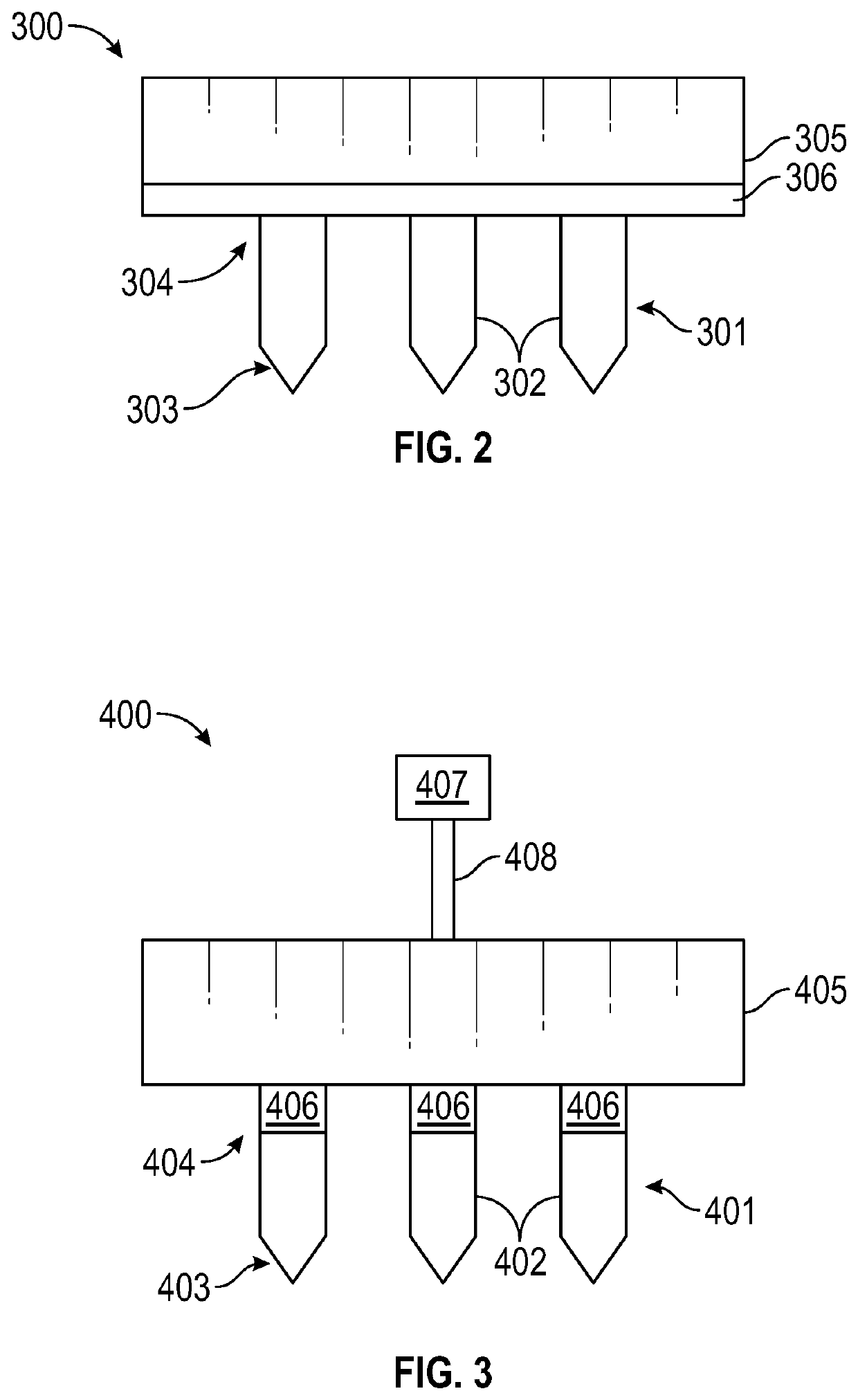
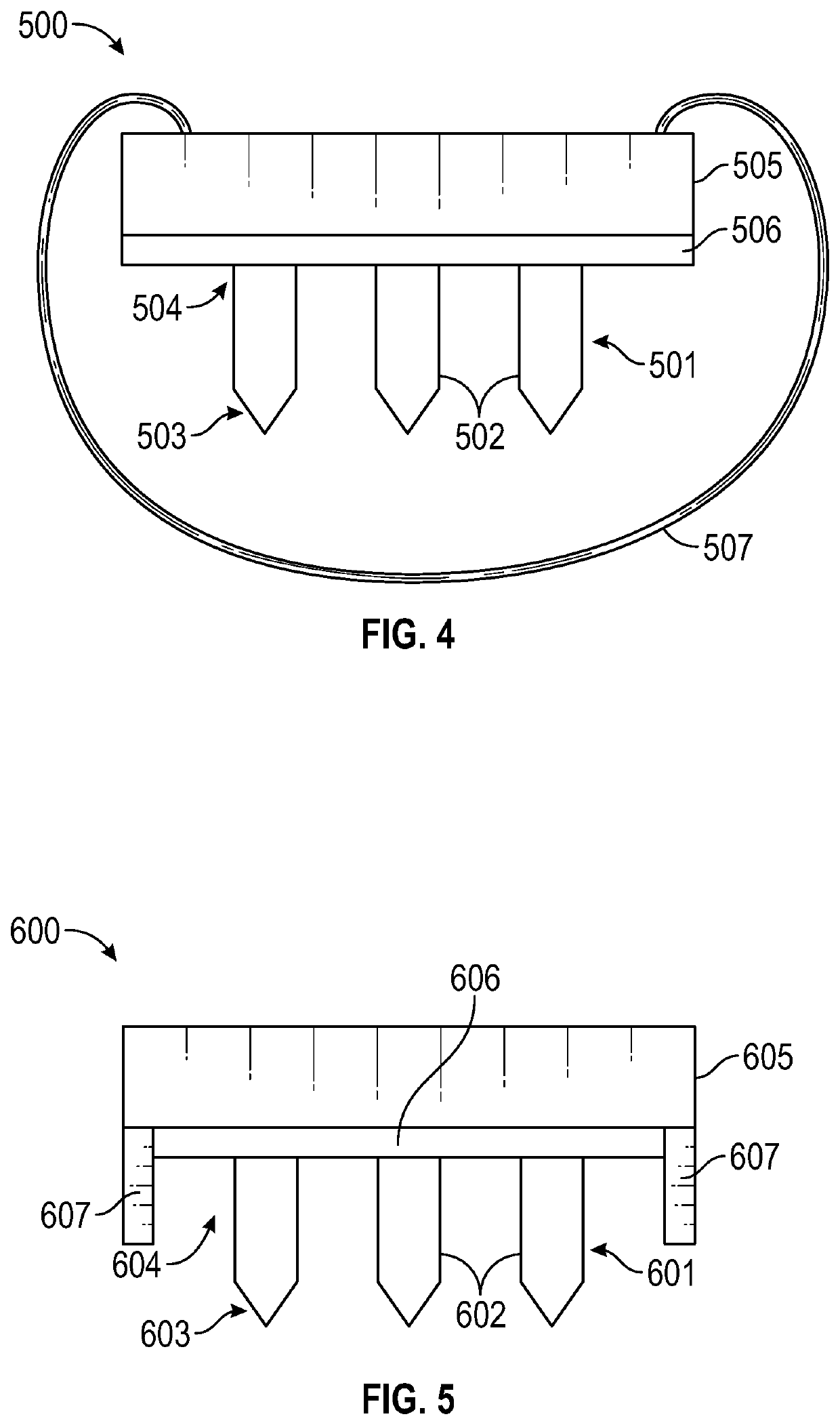
[0122]In this example, MN patches were designed in SOLIDWORKS® 2016 software (SOLIDWORKS®, Waltham, Mass.) and fabricated from grade 316 stainless steel as five-needle planar arrays (Tech-Etch, Plymouth, Mass.) by photo etching. The MN length in this example varied from 250 μm to 650 μm, with a cross-sectional area of 200 μm by 25 μm at the base and a sharp tapered tip of 10 μm diameter.

[0123]The MN patches were disinfected by washing with sterile 70% isopropyl alcohol (VWR, Radnor, Pa.) in a class II BSL hood (Thermo Fisher Scientific, Waltham, Mass.), packaged into self-sealing sterilization pouches (Crosstex International, Englewood, Colo.) and sterilized using an ethylene oxide sterilization cycle (AN74i Anprolene Gas Sterilizer, Andersen Products, Haw River, N.C.) that was validated using a product immersion method (WuXi AppTec, Marietta, Ga.).

example 2

ing Using Microneedle Patches

[0124]In this example, the MN patches of Example 1 were used to collect relatively large quantities of ISF (i.e., ≥1 μL) with a minimally invasive method that was well-tolerated, rapid, and relatively simple. Moreover, the sample of this method used equipment that is commercially available at relatively low cost.

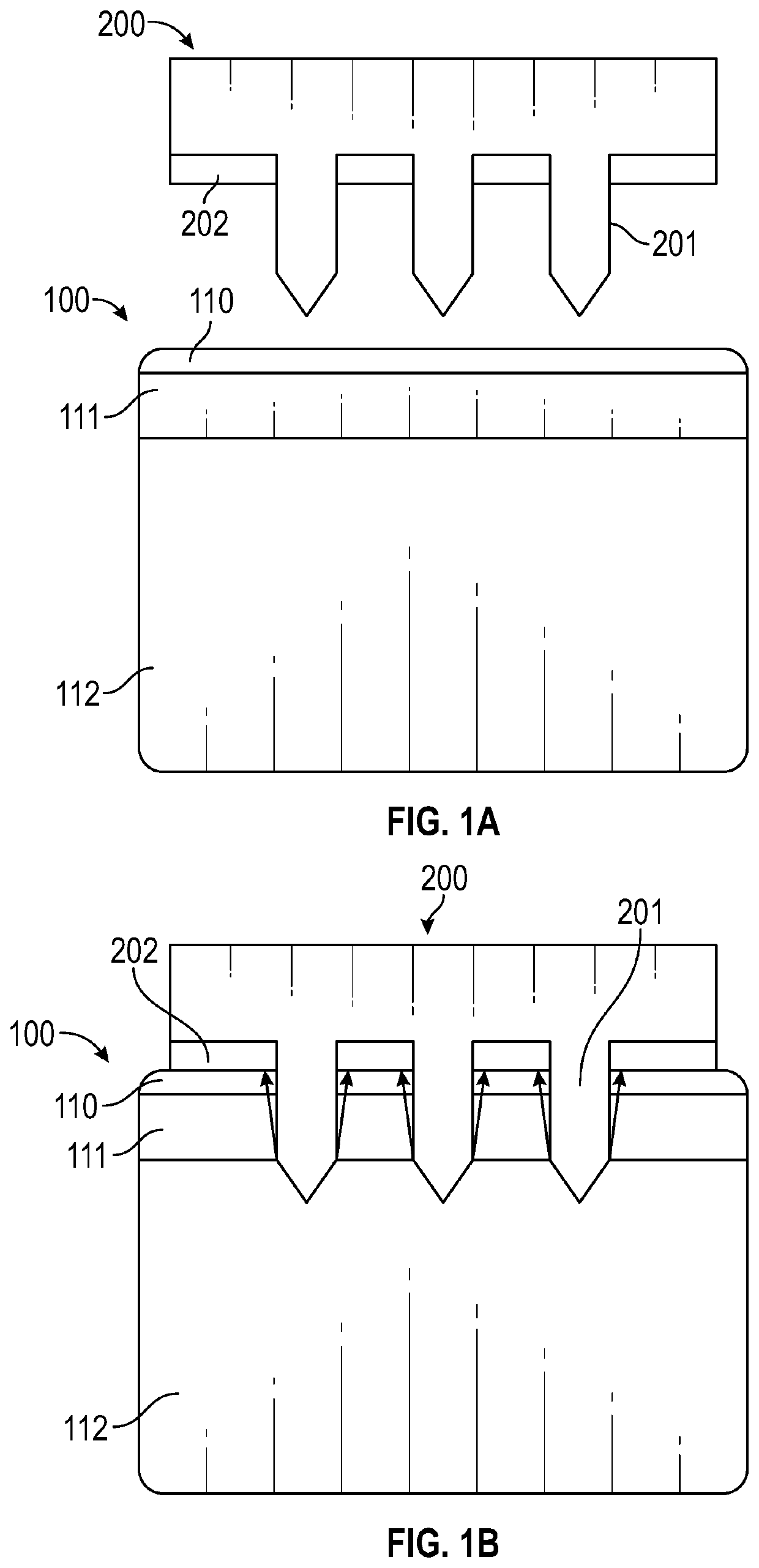
[0125]The ISF collection method of this example used MNs to create pathways for ISF flow from the skin using vacuum as a convective driving force. The vacuum application initiated a convective driving force that moved ISF through the dermis to the skin surface through MN-generated pathways. These micropores were created by pressing the patch of Example 1 into skin and then removing it. Vacuum was applied over the micropores to transport ISF through dermis and micropores to the skin surface.

[0126]FIG. 11A, FIG. 11B, FIG. 11C, and FIG. 11D are schematics depicting the method of this example. FIG. 11A depicts a sample of skin 100 that includes the s...

example 3

of Biomarkers

[0149]In this example, ISF, SBF and plasma were collected using the methods of Example 2 from 20 human participants, and more than 10,000 features were found by LC-MS analysis, more than two-thirds of which were common to ISF and plasma. This indicated that ISF may be a surrogate for plasma, at least in some cases. More features (14%) were determined to be unique to ISF by using a C18 column compared to 1% using HILIC. It was possible that the skin contained more lipophilic molecules that were captured more effectively by C18 compared to HILIC. Altogether, the data indicated that biomarkers in ISF derive mostly from plasma, there were also biomarkers specific to skin, many of which probably result from metabolic processes in the tissue.

[0150]Using a targeted approach to detect clinically valuable biomarkers, it was found that most biomarkers detected in plasma were also in ISF, further supporting the conclusion that ISF may be a substitute for blood for diagnostic tests...


PUM


Abstract

Methods and devices of collecting ISF from skin. The methods may include inserting an array of microneedles into a patients skin to form apertures in the patients skin, facilitating ISF mobilization within the skin, and collecting ISF that flows from the apertures. The devices may include an array of microneedles extending from a backing structure. The devices also may include a collection matrix for collecting ISF and/or means for facilitating ISF mobilization.

Description

CROSS-REFERENCE TO RELATED APPLICATIONS[0001]This application claims priority to U.S. Provisional Patent Application No. 62 / 741,591, filed Oct. 5, 2018, U.S. Provisional Patent Application No. 62 / 623,662, filed Jan. 30, 2018, and U.S. Provisional Patent Application No. 62 / 608,865, filed Dec. 21, 2017, which are incorporated herein by reference.BACKGROUND[0002]Biomarkers are molecules that can provide information regarding physiological status, and are found in body fluids, such as blood, urine, and saliva. Diagnosis and monitoring can be an important aspect of effective healthcare (e.g., diabetes management by blood glucose testing, quick detection of infectious diseases like HIV, and / or the ability to characterize cancers by blood sampling). Blood monitoring, however, is limited by the need for expert training, the one or more difficulties associated with continuous monitoring, the pain and / or apprehension associated with drawing blood, and the limitation of only being able to assa...

Claims


Application Information

Patent Timeline
Patent Type & Authority Applications(United States)
IPC IPC(8): A61B5/145A61B10/00
CPCA61M2037/0046A61B5/14514A61B2010/008A61M37/0015A61B10/0045A61M2037/0061A61B5/150022A61B5/150068A61B5/150099A61B5/150748A61B5/150984A61B18/00A61B17/205A61B2018/143A61M2037/0023
Inventor SAMANT, PRADNYAKOLLURU, CHANDANAPRAUSNITZ, MARK R.
Owner GEORGIA TECH RES CORP
Why Eureka
Try Eureka