Noninvasive imaging of focal atherosclerotic lesions using fluorescence molecular tomography

a fluorescence molecular tomography and focal atherosclerosis technology, applied in the field of oligopeptides, can solve the problems of rapid death, modalities not suited to frequent monitoring or even screening of at-risk patients

Inactive Publication Date: 2020-10-22
WASHINGTON UNIV IN SAINT LOUIS
View PDF0 Cites 0 Cited by
  • Summary
  • Abstract
  • Description
  • Claims
  • Application Information

AI Technical Summary

Problems solved by technology

Soft plaque can rupture and such ruptures can lead to rapid death.
Although positron emission tomography (PET) and magnetic resonance imaging (MRI) are routinely used for metabolic and morphologic imaging, these modalities are not suited for frequent monitoring or even screening of at-risk patients because of ionizing radiation (PET) and expense (PET, MRI).

Method used


Image

  • Noninvasive imaging of focal atherosclerotic lesions using fluorescence molecular tomography
  • Noninvasive imaging of focal atherosclerotic lesions using fluorescence molecular tomography
  • Noninvasive imaging of focal atherosclerotic lesions using fluorescence molecular tomography

Examples


example 1

ve Imaging of Focal Atherosclerotic Lesions Using Fluorescence Molecular Tomography

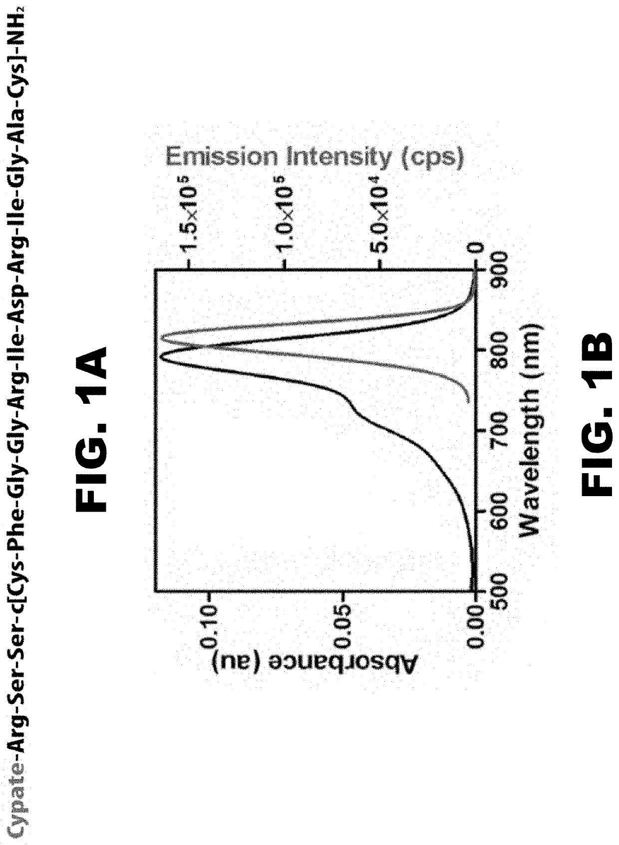
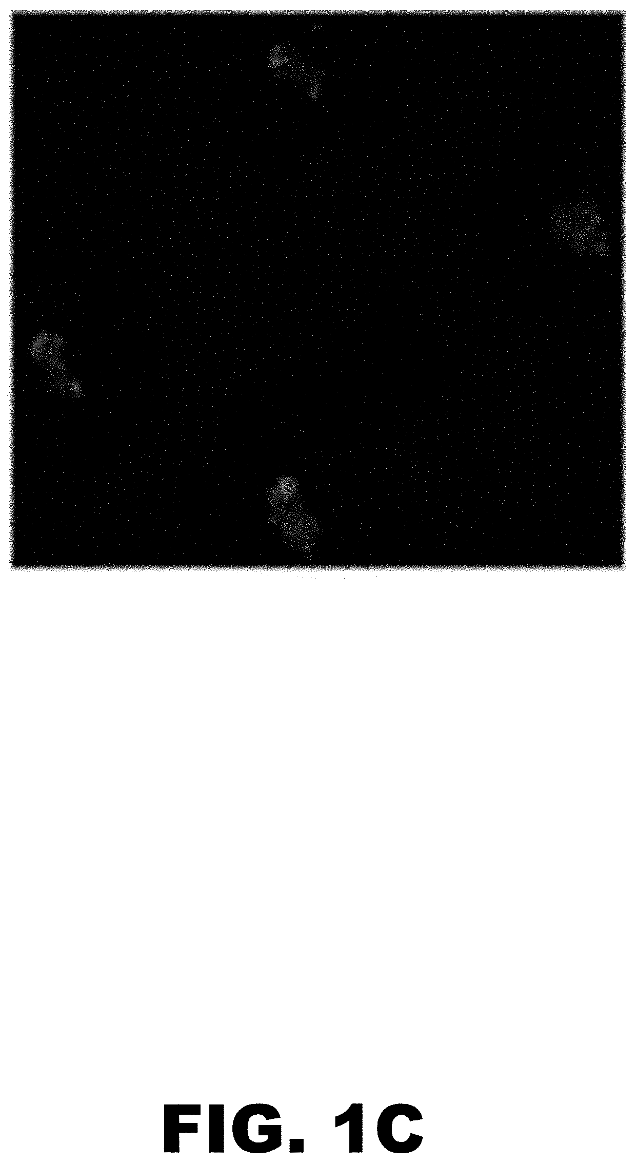
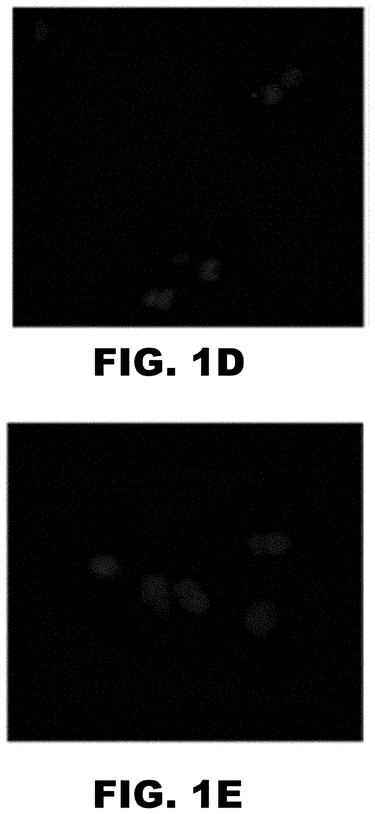
[0099]Carotid artery atherosclerosis is classified as an important cause of stroke. Unstable plaque is characterized by an eccentric neo-intimal lesion with a lipid core covered by a thinning cap of smooth muscle cells, active angiogenesis, increased matrix metalloproteinase activity, and translocation of monocyte / macrophages that transform into foam cells. Timely noninvasive imaging that could signal prerupture plaque progression will reduce the morbidity and mortality by allowing early intervention.1 Although positron emission tomography (PET) and magnetic resonance imaging (MRI) are routinely used for metabolic and morphologic imaging, these modalities are not suited for frequent monitoring or even screening of at-risk patients because of ionizing radiation (PET) and expense (PET, MRI). Transcutaneous Doppler and intravascular ultrasound are insensitive to the subtle molecular changes of critical i...


PUM

PropertyMeasurementUnit
atomic numberaaaaaaaaaa
sizeaaaaaaaaaa
lengthaaaaaaaaaa

Abstract

The present disclosure provides compositions comprising an oligopeptide comprising a fragment of a natriuretic peptide, wherein the fragment comprises the sequence Arg-Ile-Asp-Arg-Ile (SEQ ID NO:1), and a detectable label. Further disclosed are methods of imaging atherosclerotic plaque by optical imaging using a peptide composition.

Description

CROSS REFERENCE TO RELATED APPLICATIONS[0001]This application is a continuation of U.S. patent application Ser. No. 15 / 287,316, filed Oct. 6, 2016, which claims the benefit of U.S. Provisional Application No. 62 / 237,937, filed Oct. 6, 2015, the disclosures of which are hereby incorporated by reference in their entirety.GOVERNMENTAL RIGHTS[0002]This invention was made with government support under HHSN268201000046C awarded by the NIH. The government has certain rights in the invention.FIELD OF THE INVENTION[0003]The present disclosure provides compositions comprising an oligopeptide comprising a fragment of a natriuretic peptide, wherein the fragment comprises the sequence Arg-Ile-Asp-Arg-Ile (SEQ ID NO:1), and a detectable label. Further disclosed are methods of imaging atherosclerotic plaque by optical imaging using a peptide composition.BACKGROUND OF THE INVENTION[0004]Cardiovascular disease is the leading cause of death worldwide despite primary and second prevention. Each year, ...

Claims


Application Information

Patent Timeline
Patent Type & Authority Applications(United States)
IPC IPC(8): A61K49/00
CPCA61K49/0032A61K49/0056
Inventor WOODARD, PAMELA K.SHOKEEN, MONICAACHILEFU, SAMUEL
Owner WASHINGTON UNIV IN SAINT LOUIS
Why Eureka
Try Eureka