Method for noninvasive imaging of cardiac electrophysiological based on low rank and sparse constraints

a non-invasive, sparse constraint technology, applied in the field of cardiac electrophysiological analysis, can solve the problems of unable to accurately locate the specific location and shape of the focal point, obvious defects in electrocardiography, and subject risk, etc., to achieve accurate and intuitive location, and important practical value

Active Publication Date: 2020-11-10
ZHEJIANG UNIV
View PDF1 Cites 0 Cited by
  • Summary
  • Abstract
  • Description
  • Claims
  • Application Information

AI Technical Summary

Benefits of technology

[0006]The present invention provides a method for noninvasive imaging of cardiac electrophysiological based on low rank and sparse constraints. By utilizing the Spatial and temporal correlation of the distribution of ECG, this method decomposes the spatiotemporal distribution image of the endocardial and epicardial potential of the heart into a low-rank matrix representing a smooth background and a sparse matrix representing outstanding details, thereby overcoming the physical morbidity of the inverse problem of the electrocardiogram and constraining the solution to a unique optimal estimate.
[0022]In step (4), the specific method for preprocessing the surface potential data is as follows: First, the body surface potential data is denoised by means of Fourier transform or wavelet transform, and the main information in the potential signal is preserved to eliminate redundant interference. Then, the smoothing is performed on all potential signals denoised in the cardiac cycle to pull the signal baseline horizontally, avoiding the baseline deviation of the different periodic signals and introducing errors.
[0028]The present invention is based on low rank and sparse constrained non-invasive ECG imaging method, and constrains the temporal and spatial distribution of the endocardial and epicardial potential of the heart through two prior conditions of background smoothness and detail sparsity of the ECG, thereby reconstructing an accurate ECG space-time Distribution, through the spatial distribution and timing changes of the potential in the three-dimensional heart model, can accurately and intuitively obtain the location, shape and size of the lesion area, which has important practical value.

Problems solved by technology

The invasive method can provide accurate information about the internal structure and potential of the heart, but there is a certain risk in the subject during the operation.
However, there are obvious defects in electrocardiography.
The change of body surface potential is only a rough mapping of cardiac electrophysiological activity that doctors can only infer subjectively based on experience, and cannot accurately locate the specific location and shape of the focal point.
However, the above methods are based on a single prior condition (such as the smoothness of the endocardial and epicardial potential) to constrain the solution, and only utilizes the spatial distribution characteristics of the single frame potential signal.
However, the lesioned heart has more complex spatial and temporal distribution characteristics, and a single spatial smoothness constraint has certain limitations.
Therefore, it is a very significant research issue to explore a more complex prior condition of cardiac electrophysiology and utilize the spatio-temporal distribution characteristics of endocardial and epicardial potential to reconstruct the distribution of the heart potential in accordance with the real situation.

Method used


Image

  • Method for noninvasive imaging of cardiac electrophysiological based on low rank and sparse constraints
  • Method for noninvasive imaging of cardiac electrophysiological based on low rank and sparse constraints
  • Method for noninvasive imaging of cardiac electrophysiological based on low rank and sparse constraints

Examples


Embodiment Construction

[0039]In order to more specifically describe the present invention, the technical solutions of the present invention will be described in detail below in conjunction with the accompanying drawings and specific embodiments.

[0040]The present invention is based on a low rank and sparse constraint non-invasive ECG imaging method, and the specific implementation steps are as follows:

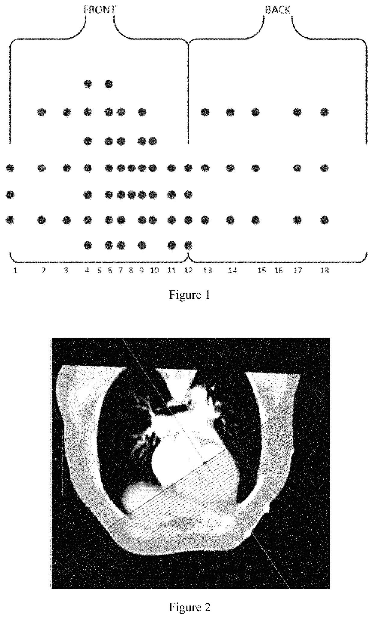
[0041]S1. Collecting the subject's 64-lead ECG and chest CT images.

[0042]First, we let the subject put on a 64-lead vest and collected the subject's 64-lead electrocardiogram to record the surface potential data of multiple cardiac cycles. The distribution of 64 leads on the body surface is shown in FIG. 1. Then, we removed the wire on the vest and retained the detection electrode only. We performed a computed tomography scan of the human chest and acquired enhanced CT image data of the subject's trunk and heart. The subject's personalized torso was recorded with the heart geometry and spatial location.

[0043]...


PUM


Abstract

The present invention discloses a method for noninvasive imaging of cardiac electrophysiological based on low rank and sparse constraints. This method decomposes the spatio-temporal distribution of endocardial and epicardial potentials into a low-rank matrix representing smooth potential components and a sparse matrix representing the details of potential salience according to the prior condition of spatio-temporal correlation of the endocardial and epicardial potential distribution of the heart. By introducing low rank and sparse constraints, the solution of the ill-conditioned inverse problem of ECG is constrained to the unique optimal solution. The invention combines the individualized three-dimensional heart model of the subject to obtain a three-dimensional dynamic distribution image of the cardiac endocardial and epicardial potential of the subject, which has important practical application value.

Description

[0001]This application claims the priority benefit of Chinese Application No. 201810027389.7, filed Jan. 11, 2018, which is hereby incorporated by reference.TECHNICAL FIELD[0002]The present invention belongs to the technical field of cardiac electrophysiological analysis, and in particular relates to a non-invasive cardiac electrophysiological inversion method based on a low-rank sparse constraint.BACKGROUND OF THE INVENTION[0003]With the continuous development of the medical level, a large number of heart disease diagnosis methods have been proposed and applied to clinical diagnosis, which are mainly divided into intrusive and non-invasive two categories. The most commonly used invasive cardiac electrophysiological imaging techniques include the Ensite system and the CARTO system. Among them, the CARTO system performs 3D electroanatomic mapping of the heart through magnetic field localization technology. The Ensite system detects the intraluminal electric field through the intralum...

Claims


Application Information

Patent Timeline
Patent Type & Authority Patents(United States)
IPC IPC(8): A61B5/00G06F17/14A61B5/0408A61B5/04G06N7/00A61B5/05
CPCA61B5/04007A61B5/0035G06F17/148A61B5/0044G06N7/00A61B5/04085A61B2576/023A61B2562/046A61B5/05A61B5/6804A61B5/7225A61B5/318G16H30/40A61B5/282G06N5/01A61B5/243
Inventor LIU, HUAFENGFANG, LIN
Owner ZHEJIANG UNIV
Why Eureka
Try Eureka