Expansion Of Peripheral Blood Lymphocytes (PBLS) From Peripheral Blood

a peripheral blood and lymphocyte technology, applied in the field of peripheral blood lymphocyte expansion from peripheral blood, can solve the problems of poor results of earlier approaches to expansion of tils from b cell lymphomas, and the field remains challenging

Inactive Publication Date: 2020-11-05
IOVANCE BIOTHERAPEUTICS INC
View PDF2 Cites 22 Cited by
  • Summary
  • Abstract
  • Description
  • Claims
  • Application Information

AI Technical Summary

Problems solved by technology

A number of approaches to improve responses to TIL therapy in melanoma and to expand TIL therapy to other tumor types have been explored with limited success, and the field remains challenging.
Earlier approaches to expansions of TILs from B cell lymphomas yielded poor results, with only 2 of 12 attempts at TIL growth providing for potential activity against tumors.

Method used


Image

  • Expansion Of Peripheral Blood Lymphocytes (PBLS) From Peripheral Blood
  • Expansion Of Peripheral Blood Lymphocytes (PBLS) From Peripheral Blood
  • Expansion Of Peripheral Blood Lymphocytes (PBLS) From Peripheral Blood

Examples


example 1

Expansion of TILs from Non-Hodgkin's Lymphomas

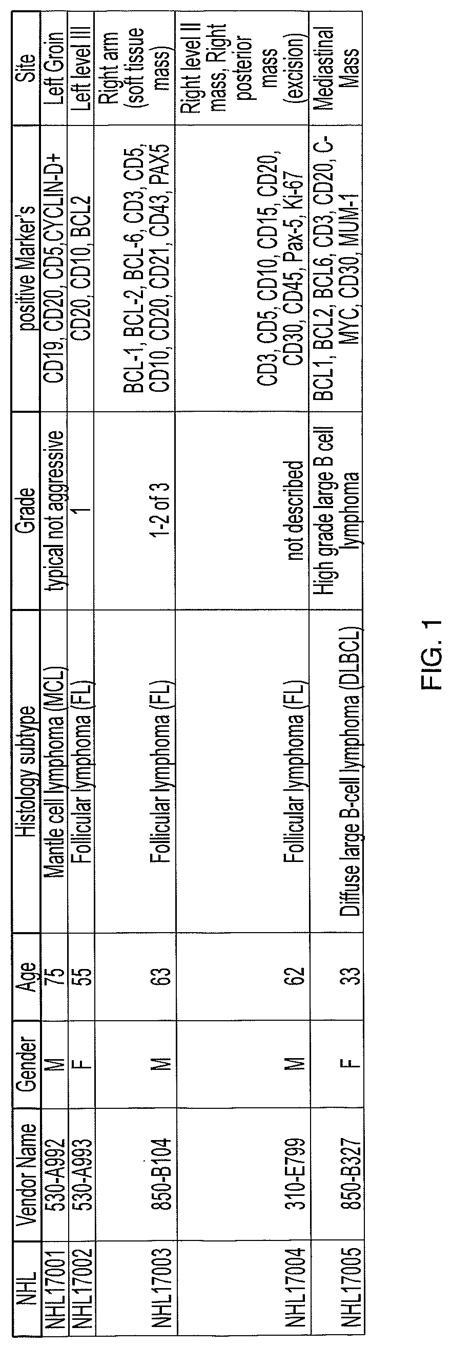
[0595]TILs were expanded from five non-Hodgkin's lymphoma tumors (one mantle cell lymphoma tumor, three follicular lymphoma tumors, and one ABC-type diffuse large B cell lymphoma tumor) with the pathologies given in FIG. 1, using IL-2 for 11 to 14 days in a pre-REP stage, followed by subsequent REP for 14 days using IL-2, mitogenic anti-CD3 antibody, and irradiated allogeneic peripheral blood mononuclear cell (PBMC) feeders. TILs were successfully generated from all 5 lymphoma tumors with maximum expansion index of 680 fold, significantly higher than previously observed using other methods. Schwartzentruber, et al., Blood 1993, 82, 1204-1211. Further, mean CD3+ T cell population was 95% (versus 75% using the method of Schwartzentruber, et al., Blood 1993, 82, 1204-1211).

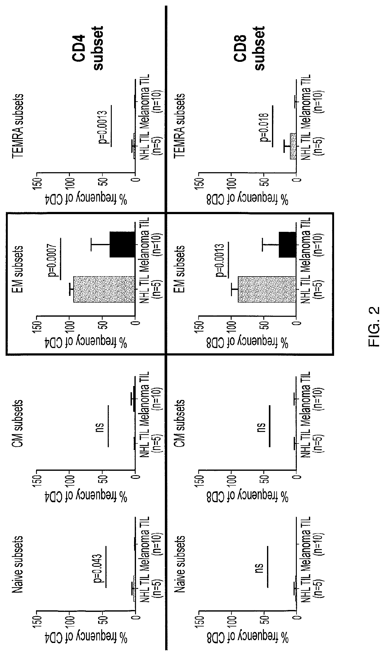
[0596]Cell sorting and flow cytometry was performed using a Becton, Dickinson & Co. (BD) FACS CANTO II system. A marked relative increase in effector memory cells that wa...

example 2

Phenotypic and Functional Characterization of Marrow Infiltrating Lymphocytes (MIL) Grown from Bone Marrow of AML Patients and Peripheral Blood Lymphocytes (PBL) Grown from Peripheral Blood of AML Patients

[0603]Samples of bone marrow and as available a related blood sample were obtained from patients with acute myeloid leukemia (AML), including patients pre-treated with at least three rounds of a regimen comprising ibrutinib (1-[(3R)-3-[4-amino-3-(4-phenoxyphenyl)-1H-pyrazolo[3,4-d]pyrimidin-1-yl]-1-piperidinyl]-2-propen-1-one), accompanied by information about the patient's age, gender, stage, tumor type, site of cancer, treatment history, a de-identified pathology report, and any molecular tests performed (e.g., MSI expression and Raf / Ras expression). MILs and PBLs were expanded using one of MIL Method 1, MIL Method 2, or MIL Method 3, or PBL Method 1, PBL Method 2, or PBL Method 3, and the MILs and PBLs were phenotypically and functionally characterized.

[0604]FIGS. 36A and 36B il...

example 3

Methods of Expanding TILs and Treating Cancers with Expanded TILs

[0612]Bone marrow is obtained using needle aspiration. The bone marrow sample is aspirated into heparin-containing syringes and stored overnight at room temperature. After storage, the contents of the syringes are pooled together into a sterile container and quality tested. The bone marrow is enriched for mononuclear cells (MNCs) using lymphocyte separation media (LSM) and centrifugation with a COBE Spectra. Cells in the gradient are collected down to the red blood cells and washed using HBSS. The MNCs are cryopreserved using a hetastarch-based cryoprotectant supplemented with 2% HSA and 5% DMSO, reserving some of the MNCs for quality control. The QC vial is thawed to determine the CD3+ and CD38+ / 138+ cell content of the MNC product.

[0613]The bone marrow is aspirated and fractionated on a Lymphocyte Separation Medium density gradient and cells are collected almost to the level of the red cell pellet. This fractionation...


PUM

PropertyMeasurementUnit
timeaaaaaaaaaa
molecular weightaaaaaaaaaa
molecular massaaaaaaaaaa

Abstract

Methods of expanding tumor infiltrating lymphocytes (TILs), including peripheral blood lymphocytes (PBLs) and marrow infiltrating lymphocytes (MILs), from blood and / or bone marrow of patients with hematological malignancies, such as liquid tumors, including lymphomas and leukemias, and genetic modifications of expanded TILs, PBLs, and MILs to incorporate chimeric antigen receptors, genetically modified T-cell receptors, and other genetic modifications, and uses of such expanded and / or modified TILs, PBLs, and MILs in the treatment of diseases such as cancers and hematological malignancies are disclosed herein.

Description

CROSS REFERENCE TO RELATED APPLICATIONS[0001]This application is a continuation-in-part of International Patent Application No. PCT / US18 / 032109 filed May 10, 2018, which claims priority to U.S. Provisional Application No. 62 / 504,337, filed May 10, 2017; U.S. Provisional Application No. 62 / 530,681, filed Jul. 10, 2017; U.S. Provisional Application No. 62 / 550,398, filed Aug. 25, 2017; U.S. Provisional Application No. 62 / 590,034, filed Nov. 22, 2017; U.S. Provisional Application No. 62 / 621,462, filed Jan. 24, 2018; U.S. Provisional Application No. 62 / 621,798, filed Jan. 25, 2018; and U.S. Provisional Application No. 62 / 647,367, filed Mar. 23, 2018, all of which are herein incorporated by reference in their entireties.FIELD OF THE INVENTION[0002]Methods of expanding tumor infiltrating lymphocytes (TILs) derived from blood and / or bone marrow of a patient with a hematological malignancy, such as a liquid tumor, including lymphomas and leukemias, and compositions comprising populations of ...

Claims


Application Information

Patent Timeline
Patent Type & Authority Applications(United States)
IPC IPC(8): C12N5/0783A61K35/17
CPCA61K35/17C12N5/0636C12N2501/51C12N2502/1107C12N2501/515C12N2501/2302C12N5/0635C12N5/0638C12N2502/11C12N2502/30A61P35/00A61K31/519A61K38/2013C12N5/0634C12N2500/00C12N2501/998C12N2501/999
Inventor KARYAMPUDI, LAVAKUMARFARDIS, MARIA
Owner IOVANCE BIOTHERAPEUTICS INC
Why Eureka
Try Eureka