Expansion microscopy compatible and multiplexed in situ hybridization of formalin fixed paraffin embedded tissue sections for spatially resolved transcriptomics

a paraffin embedded tissue and expansion microscopy technology, applied in the field of imaging, can solve the problems of reducing the ability to systematically map transcriptional variations across a large number of genes, reducing the accuracy of rna spot count quantification, and limited strategy

Active Publication Date: 2020-11-12
EXPANSION TECH
View PDF0 Cites 10 Cited by
  • Summary
  • Abstract
  • Description
  • Claims
  • Application Information

AI Technical Summary

Benefits of technology

[0010]In one aspect, provided herein are methods of labeling nucleic acids and proteins together in a formalin fixed paraffin embedded (FFPE) biological tissue sample adhered on a slide, the methods comprising: (a) performing deparaffinization and antigen retrieval on the sample; (b) contacting the sample with a first gel binding moiety and a second gel binding moiety under conditions wherein the first gel binding moiety operably links to proteins in the sample and the second gel binding moiety operably links to nucleic acids in the sample; (c) contacting the sample with a solution comprising monomers of a polyelectrolyte gel; (d) by free radical polymerization, polymerizing the monomers to form the polyelectrolyte gel and covalently conjugating the first and second gel binding moieties to the polyelectrolyte gel; (e) proteolytically digesting the sample; (f) detaching the polyelectrolyte gel from the slide; (g) dialyzing the polyelectrolyte gel to expand it; (h) providing a plurality of initiator deoxyribonucleic acid (DNA) probes targeting a plurality of nucleic acid targets of interest, wherein each of the plurality of initiator DNA probes comprise (A) a sequence complementary to a sequence from one of the nucleic acid targets of interest, and (B) a hybridization chain reaction (HCR) initiator sequence; (i) contacting the gel with the plurality of initiator DNA probes under conditions wherein the sequence complementary to the sequence from one of the nucleic acid targets of interest hybridizes to that sequence from the nucleic acid target of interest; (j) for each initiator DNA probe, providing a pair of fluorophore-labeled DNA hairpins that metastably co-exist in the absence of the initiator DNA probe; and (k) contacting the gel with the fluorophore-labeled DNA hairpins under conditions wherein the hairpins self-assemble by HCR, in the presence of their corresponding initiator DNA probe, into fluorescent amplification polymers tethered to that corresponding initiator DNA probe. In some embodiments, the methods further comprise, prior to step (h), embedding the expanded gel in a polyacrylamide gel matrix. In some embodiments, the methods further comprising, prior to step (b): (I) contacting the sample with one or more primary antibodies under conditions where they selectively recognize one or more target proteins of interest; and (II) contacting the sample with one or more secondary antibodies operably linked to a detectable label. In some embodiments, the steps of (I) contacting the sample with one or more primary antibodies under conditions where they selectively recognize one or more target proteins of interest; and (II) contacting the sample with one or more secondary antibodies operably linked to a detectable label; are repeated after the gel is detached from the slide.

Problems solved by technology

However the analysis of ribonucleic acid (RNA) within clinical samples is currently constrained by the diffraction limit of conventional microscopes which reduce the accuracy of RNA spot count quantification.
Additionally, the limited number of transcripts (typically 3-4 depending on the number of available fluorescent channels) further reduces the ability to systematically map transcriptional variations across a large number of genes.
However, because the number of fluorescent dyes that can be used is small (typically <6), this strategy is limited to imaging only a small number of proteins per sample.
Additionally, the polymerization process dampens the fluorescence of the dyes, which are permanently connected to the gel matrix.
Also, the presence of the DNA on the antibody reduces the extent and the rate at which it binds to the target.
The result is that the current ExM processes are lengthy, and the staining is commonly dim compared to controls, making this approach laborious, time consuming, and inefficient for RNA detection due to the limited number of transcripts which can be resolved within a single specimen.
However, this approach is suitable for protein targets but not RNA detection.
The interrogation of RNA in clinical samples in a highly multiplexed and efficient manner remains a challenge in structural biology and medicine.

Method used


Image

  • Expansion microscopy compatible and multiplexed in situ hybridization of formalin fixed paraffin embedded tissue sections for spatially resolved transcriptomics
  • Expansion microscopy compatible and multiplexed in situ hybridization of formalin fixed paraffin embedded tissue sections for spatially resolved transcriptomics
  • Expansion microscopy compatible and multiplexed in situ hybridization of formalin fixed paraffin embedded tissue sections for spatially resolved transcriptomics

Examples


example 1

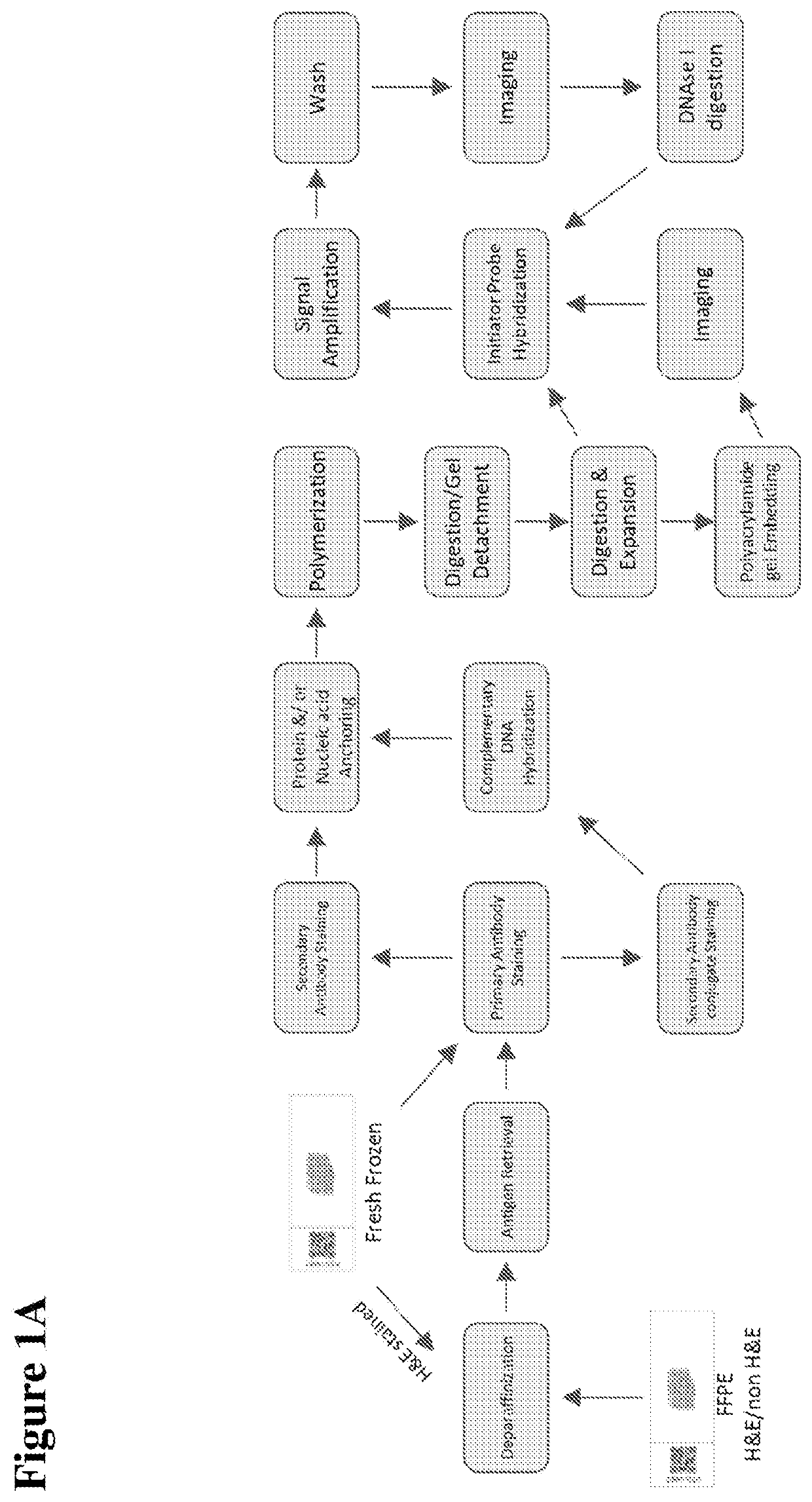
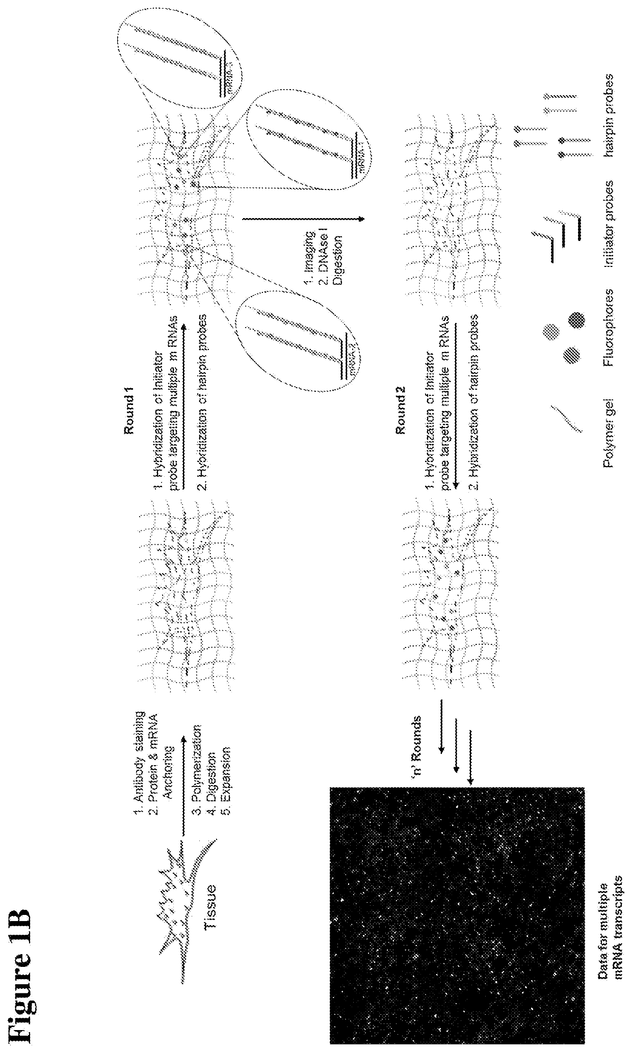
[0177]As shown in FIGS. 1A and 1B, and an FFPE biological sample adhered on a slide (e.g., a frosted glass slide) is obtained. Under RNAse-free conditions, the sample undergoes deparaffinization, followed by antigen retrieval and subsequent staining with primary and secondary antibodies. Also under RNAse-free conditions, the RNA in the sample is linked, directly or indirectly, to a gel binding moiety. The sample is then contacted with a solution comprising monomers of a polyelectrolyte gel, which are then polymerized by free radical polymerization to form the polyelectrolyte gel, as well as anchoring the protein and RNA to the gel. The sample is digested, and the gel is detached from the slide; the gel is then dialyzed to expand it, optionally followed by polyacrylamide embedding and imaging. The next step is probe hybridization, HCR amplification, followed by washing, imaging, and digestion with DNAse I. Subsequent rounds of probe, hybridization, HCR amplification, washing, imaging...

example 2

[0178]More specifically, for an RNA target of interest (e.g., an mRNA), a single-stranded DNA probe is provided having a 5′ sequence complementary or partially complementary to a sequence of the RNA target of interest, and a 3′ HCR initiator domain having a first initiator segment and a 3′ second initiator segment. The probe is optionally also operably linked to a detectable label. The expanded gel is contacted with the DNA probe under conditions to selectively hybridize with the RNA target of interest. If the probe includes a detectable label, an image may be taken of the gel.

[0179]An HCR amplifier is provided, which comprises a first DNA hairpin molecule and a second DNA hairpin molecule, and which coexist metastably in the absence of the probes hybridized to the RNA target of interest. The first DNA hairpin molecule sequentially comprises (i) a first domain comprising a 5′ tail complementary or partially complementary to the 3′ second initiator segment of the 3′ HCR intiator doma...

example 3

[0183]For an RNA target of interest (e.g., an mRNA) in a biological sample of interest, and a plurality of unique single-stranded DNA probes are provided. Each unique probe has a 5′ sequence complementary or partially complementary to a unique sequence of mRNA target of interest, and a 3′ HCR initiator domain having a first initiator segment and a 3′ second initiator segment The probes are optionally also operably linked to a detectable label. The expanded sample is contacted with the DNA probes under conditions to selectively hybridize with the RNA targets of interest. If the probes include a detectable label, an image may be taken of the gel.

[0184]An HCR amplifier is provided. The HCR amplifier comprises a first DNA hairpin molecule and a second DNA hairpin molecule, which coexist metastably in the absence of the probes hybridized to the RNA target of interest. The first DNA hairpin molecule sequentially comprises (i) a first domain comprising a 5′ tail complementary or partially ...


PUM

PropertyMeasurementUnit
pHaaaaaaaaaa
dissociation constantaaaaaaaaaa

Abstract

The invention relates to imaging, such as in situ imaging by expansion microscopy, labelling, and analyzing biological samples, such as formalin fixed paraffin embedded (FFPE) cells and tissues, as well as reagents and kits for doing so.

Description

FIELD OF THE INVENTION[0001]The invention relates to imaging, such as in situ imaging by expansion microscopy, labelling, and analyzing biological samples, such as formalin fixed paraffin embedded (FFPE) cells and tissues, as well as reagents and kits for doing so.BACKGROUND OF THE INVENTION[0002]In situ transcriptional profiling of clinical formalin fixed paraffin embedded (FFPE) tissues has the potential to contribute to the development of accurate and reliable diagnoses for diseases, such as cancer. However the analysis of ribonucleic acid (RNA) within clinical samples is currently constrained by the diffraction limit of conventional microscopes which reduce the accuracy of RNA spot count quantification. Additionally, the limited number of transcripts (typically 3-4 depending on the number of available fluorescent channels) further reduces the ability to systematically map transcriptional variations across a large number of genes.[0003]Current imaging technologies for analyzing R...

Claims


Application Information

Patent Timeline
Patent Type & Authority Applications(United States)
IPC IPC(8): C12Q1/6841G01N1/30
CPCC12Q2527/125C12Q1/6841C12Q2525/301G01N1/30C12N15/10G01N33/54306
Inventor DEWAL, MAHENDER BABU
Owner EXPANSION TECH
Why Eureka
Try Eureka