Tissue chip used for tumour early stage diagnosis and preparation device

A tissue chip and early diagnosis technology, applied in instruments, analytical materials, etc., can solve problems such as high-throughput, early diagnosis and prediction of malignant tumors that cannot be screened

Inactive Publication Date: 2004-12-22
中南大学湘雅医学院肿瘤研究所
View PDF0 Cites 19 Cited by
  • Summary
  • Abstract
  • Description
  • Claims
  • Application Information

AI Technical Summary

Problems solved by technology

[0003] The present invention aims to develop a new type of tissue chip to solve the problem that the current tissue chip

Method used


Image

  • Tissue chip used for tumour early stage diagnosis and preparation device
  • Tissue chip used for tumour early stage diagnosis and preparation device
  • Tissue chip used for tumour early stage diagnosis and preparation device

Examples


Embodiment

[0018] Example: nasopharyngeal carcinoma tissue chip and its fabrication

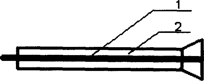
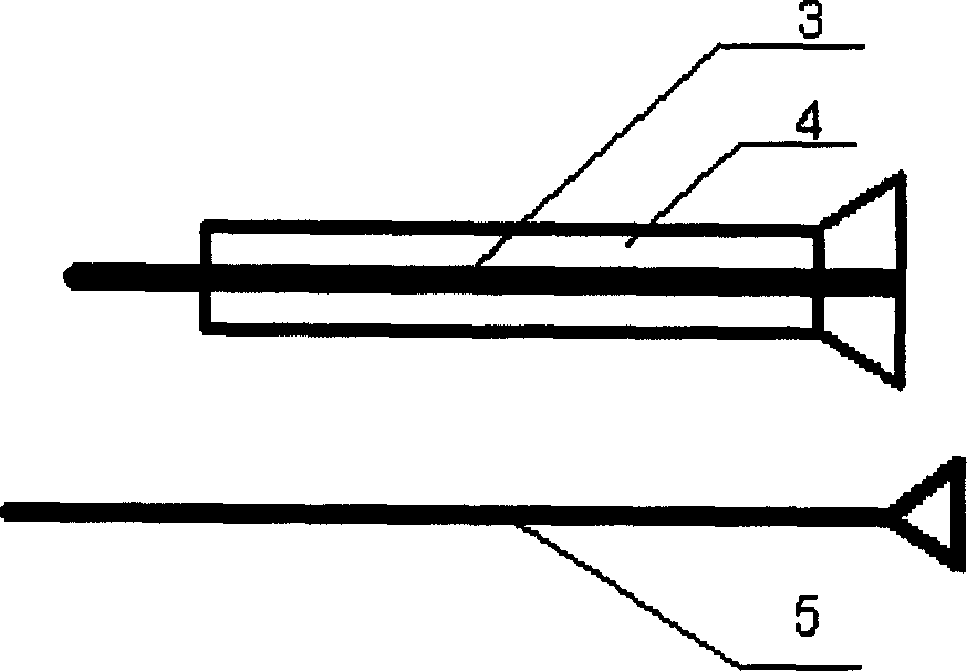
[0019] (1) Specimen preparation and target tissue HE staining section positioning: 250 paraffin tissue specimens of nasopharyngeal carcinoma, 150 cases of precancerous lesions and 48 cases of corresponding normal nasopharyngeal tissues were selected from the tissue specimen bank with complete clinical and pathological characteristics. , Routine dehydration, paraffin embedding, sectioning, HE staining, confirming the correctness of the tissue under the microscope, and marking and positioning the target tissue in the section under the microscope. (2) Production of recipient paraffin blocks: tissue microarrays with a tissue diameter of 1.0mm, including nasopharyngeal carcinoma, precancerous lesions and normal nasopharyngeal tissues, with a total of 448 specimens, using mold paper with 448 tissue point arrays ( image 3 ) on the surface of the receptor wax block to guide the preparation of the receptor hole...


PUM


Abstract

Three kinds of tissues including cancer tissue, precancerosis and corresponding normal tissue are sliced up, dyed, marked, and positioned. Receptor holes are prepared by leading designed lattice array mould paper to paste on surface of wax block of receptor. Wax block with tissue core bar is prepared by using perforating needle and puncture needle for tissue. Common cancer such as lung cancer, nasopharyngeal carcinoma, oesophagus cancer etc. and having integrated clinical data and pathology features are selected. Through in situ hybridization, testing mRNA of relevant gene and expression of protein on tissue chip, consistent result between the invented product and traditional test is validated. In the product, cellular morphology is clear and even, and there is no fallen off tissue point. The invention is applicable to filter cancers, early diagnosis and forecasting prognosis.

Description

Technical field: [0001] The invention relates to biological products for tissue detection, in particular to a tissue chip. Background technique: [0002] Tissue chip, also known as tissue microarray (Tissue chip, Tissue Microarray TMA,) is that many donor tissues are rearranged on the recipient wax block in the form of a microarray, and the recipient wax block is sliced ​​and transferred to a glass slide. Sections including hundreds of tissues can be used for a variety of in situ expression studies. Tissue microarrays are often used to puncture the tissue of the donor wax block and make the acceptor hole of the recipient wax block. At present, there are tumor tissue chips, normal tissue chips, single or composite tissue chips, etc. on the market, but they cannot be used for high-throughput screening of molecular markers for early diagnosis of malignant tumors and prediction of prognosis. Therefore, it is urgent to make a tissue microarray that can be used to detect the exp...

Claims


Application Information

Patent Timeline
IPC IPC(8): G01N35/00
Inventor 李桂源范松青马键李小玲谭琛熊炜黄河周鸣
Owner 中南大学湘雅医学院肿瘤研究所
Why Eureka
Try Eureka