System, method and computer-accessible medium for classifying breast tissue using a convolutional neural network

a convolutional neural network and breast tissue technology, applied in the field of breast tissue classification and classification information, can solve the problems of increasing healthcare costs, increasing physical and psychological stress on patients and their families, and difficult to distinguish from ductal carcinoma in situ, so as to limit the drift of layer activation

Inactive Publication Date: 2020-11-19
THE TRUSTEES OF COLUMBIA UNIV IN THE CITY OF NEW YORK
View PDF0 Cites 15 Cited by
  • Summary
  • Abstract
  • Description
  • Claims
  • Application Information

AI Technical Summary

Benefits of technology

[0013]In certain exemplary embodiments of the present disclosure, a score(s) can be determined based on the image(s) using the neural network(s). The breast tissue can be automatically classified based on the score (e.g., a score above 0.5). The image(s) can illustrate excised breast tissue(s). The image(s) can be segmented and resized prior to classifying the breast tissue. A batch normalization can be performed on the image(s), which can be used so as to limit a drift of layer activations.

Problems solved by technology

ADH is diagnosed by biopsy in up to 15% of suspicious screen-detected lesions, and is often difficult to distinguish from ductal carcinoma in situ (“DCIS”).
In addition, even with the use of vacuum-assisted biopsy procedures, biopsy alone has resulted in unacceptably high rates of upgrade.
Approximately 23% of patients may require surgical re-excision (see, e.g., References 26 and 27), which leads to increased healthcare costs and physical and psychological stress on patients and their families.
Interpretation of OCT images is typically performed by researchers and clinicians, but manual image interpretation is challenging due to its slow speed and time to train readers, high interobserver variability, and image complexity.
As a result, a manual interpretation is not practical in an intraoperative setting.

Method used


Image

  • System, method and computer-accessible medium for classifying breast tissue using a convolutional neural network
  • System, method and computer-accessible medium for classifying breast tissue using a convolutional neural network
  • System, method and computer-accessible medium for classifying breast tissue using a convolutional neural network

Examples


Embodiment Construction

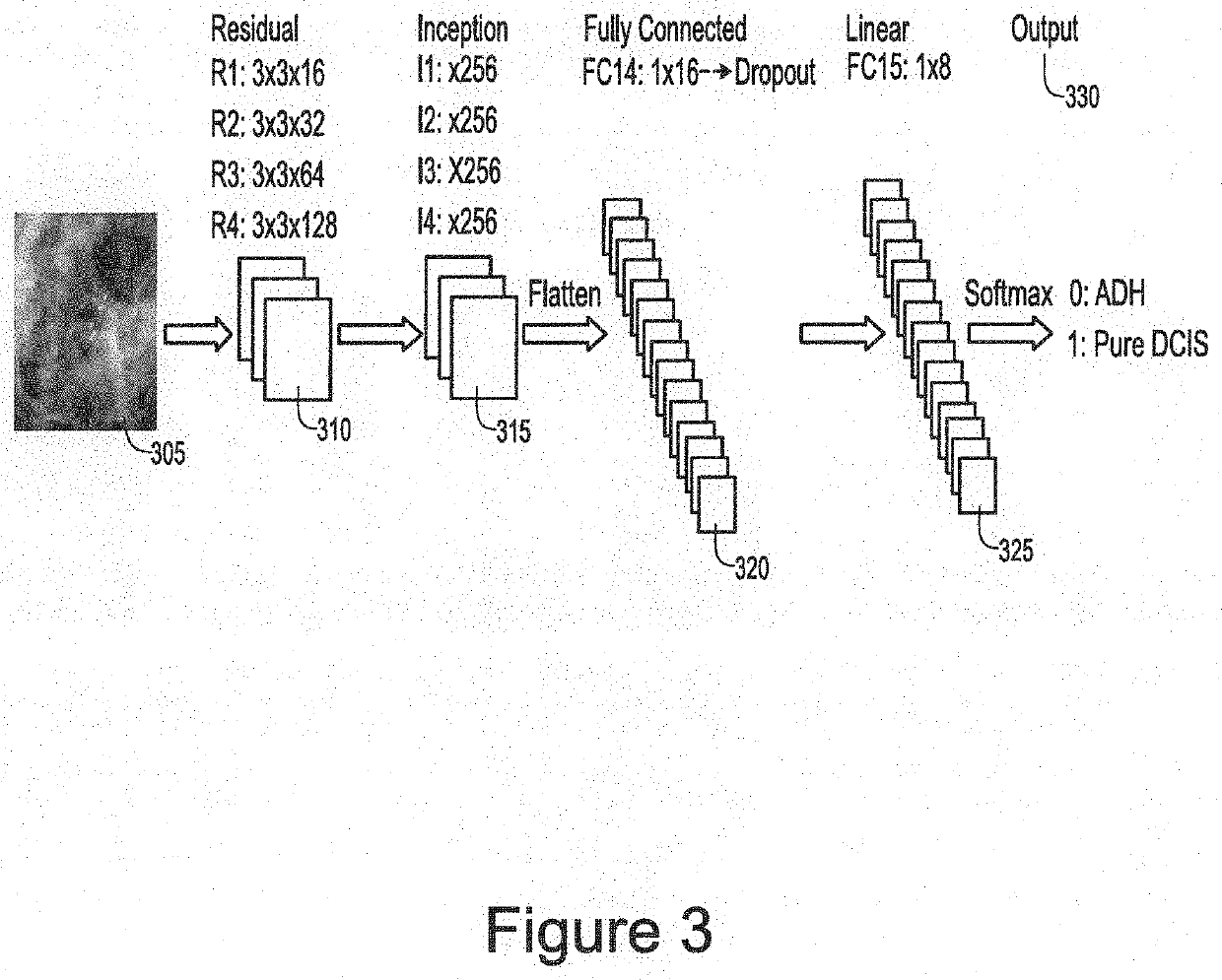
[0011]An exemplary system, method and computer-accessible medium for classifying a breast tissue(s) a patient(s) can include, for example, receiving an image(s) of an internal portion(s) of a breast of the patient(s), and automatically classifying the breast tissue(s) of the breast by applying a neural network(s) to the image(s). The automatic classification can include a classification as to whether the breast tissue(s) is atypical ductal hyperplasia or ductal carcinoma. The automatic classification can include a classification as to whether the breast tissue(s) is a cancerous tissue or a non-cancerous tissue. The image(s) can be a mammographic image or an optical coherence tomography image.

[0012]In some exemplary embodiments of the present disclosure, the neural network can be a convolutional neural network (CNN). The CNN can include a plurality of layers. The layers can include (i) a plurality of residual layers, (ii) a plurality of inception layers, (iii) a fully connected layer...


PUM


Abstract

An exemplary system, method and computer-accessible medium for classifying a breast tissue(s) a patient(s) can include, for example, receiving an image(s) of an internal portion(s) of a breast of the patient(s), and automatically classifying the breast tissue(s) of the breast by applying a neural network(s) to the image(s). The automatic classification can include a classification as to whether the breast tissue(s) is atypical ductal hyperplasia or ductal carcinoma. The automatic classification can include a classification as to whether the breast tissue(s) is a cancerous tissue or a non-cancerous tissue. The image(s) can be a mammographic image or an optical coherence tomography image.

Description

CROSS-REFERENCE TO RELATED APPLICATION(S)[0001]This application relates to and claims priority from U.S. Patent Application No. 62 / 589,924, filed on Nov. 22, 2017, the entire disclosure of which is incorporated herein by reference.FIELD OF THE DISCLOSURE[0002]The present disclosure relates generally to a classification of information regarding breasts and breast tissue, and more specifically, to exemplary embodiments of systems, methods and computer-accessible medium for classifying breast tissue using a convolutional neural network.BACKGROUND INFORMATION[0003]Atypical ductal hyperplasia (“ADH”) is a proliferative epithelial lesion involving the terminal ductal lobular units of the breast that is a non-obligate precursor to invasive disease. ADH is diagnosed by biopsy in up to 15% of suspicious screen-detected lesions, and is often difficult to distinguish from ductal carcinoma in situ (“DCIS”). (See, e.g., Reference 1). While ADH is morphologically very similar to low-grade DCIS, t...

Claims


Application Information

Patent Timeline
Patent Type & Authority Applications(United States)
IPC IPC(8): G06T7/00G06K9/62G06T11/00G06T7/10G06T3/40G06N3/08G06N3/04G16H50/20G16H30/40A61B5/00
CPCG06T2207/30068G06T2207/30096G06T2207/20084G06N3/08G06T7/0012G06K9/6279G16H50/20G06T3/4046A61B2576/02A61B5/0066G06T11/003G06T2207/10101A61B5/7264A61B5/0091G06K9/6217G06T7/10G06N3/04G16H30/40G06T2207/10088G06T2207/20081G06T2207/20132G16H50/30G06T3/40G06T3/60G06T5/50G06T2207/10096G06T2207/20224G06T7/11G06F18/21G06F18/243G06N3/045
Inventor HA, RICHARD
Owner THE TRUSTEES OF COLUMBIA UNIV IN THE CITY OF NEW YORK
Why Eureka
Try Eureka