Systems and methods for brain hemorrhage classification in medical images using an artificial intelligence network

an artificial intelligence network and brain hemorrhage technology, applied in image enhancement, tomography, instruments, etc., can solve the problems of insufficient accuracy to warrant being relied upon, deep learning routines may not be sensitive enough to distinguish between regions in images, and learning networks cannot process images at a speed that meets clinical needs

an artificial intelligence network and brain hemorrhage technology, applied in image enhancement, tomography, instruments, etc., can solve the problems of insufficient accuracy to warrant being relied upon, deep learning routines may not be sensitive enough to distinguish between regions in images, and learning networks cannot process images at a speed that meets clinical needs

US20210082566A1Active Publication Date: 2021-03-18THE GENERAL HOSPITAL CORP

Image

  • Systems and methods for brain hemorrhage classification in medical images using an artificial intelligence network
  • Systems and methods for brain hemorrhage classification in medical images using an artificial intelligence network
  • Systems and methods for brain hemorrhage classification in medical images using an artificial intelligence network

Examples


Embodiment Construction

[0017]Systems and methods for rapid, accurate, fully-automated brain hemorrhage deep learning (DL) based assessment tools are provided. These systems and methods can be used, as an example, to assist clinicians in the detection and characterization of hemorrhages. Hemorrhages may include intracranial hemorrhage (ICH), subarachnoid hemorrhage (SAH), intra-parenchymal hemorrhage (IPH), epidural / subdural hematoma (SDH), and the like.

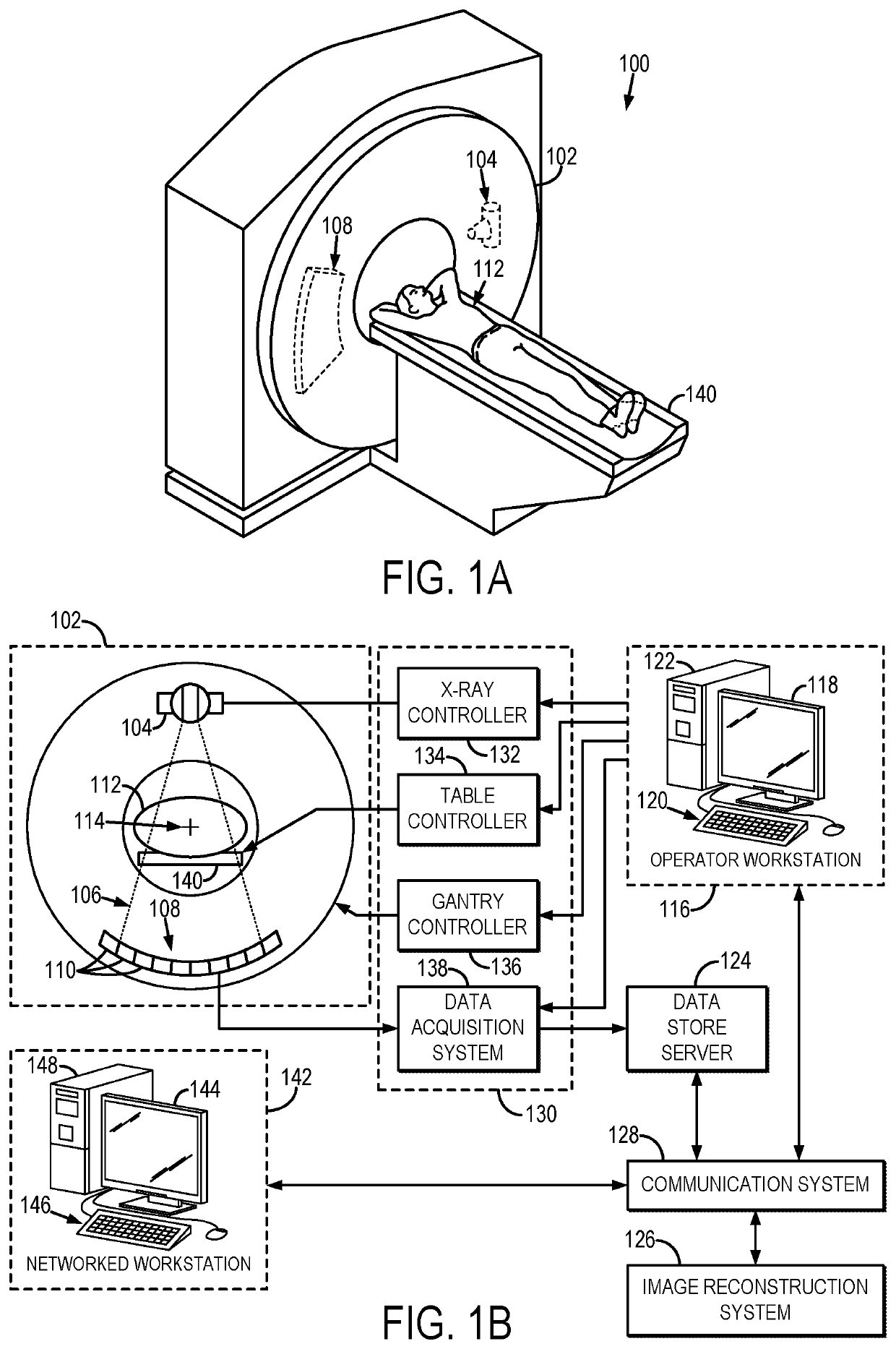
[0018]Referring particularly now to FIGS. 1A and 1B, an example of an x-ray computed tomography (“CT”) imaging system 100 is illustrated for use with some configurations of the disclosure. The CT system includes a gantry 102, to which at least one x-ray source 104 is coupled. The x-ray source 104 projects an x-ray beam 106, which may be a fan-beam or cone-beam of x-rays, towards a detector array 108 on the opposite side of the gantry 102. The detector array 108 includes a number of x-ray detector elements 110. Together, the x-ray detector elements 110 sense...


PUM


Abstract

Systems and methods for rapid, accurate, fully-automated, brain hemorrhage deep learning (DL) based assessment tools are provided, to assist clinicians in the detection & characterization of hemorrhages or bleeds. Images may be acquired from a subject using an imaging source, and preprocessed to cleanup, reformat, and perform any needed interpolation prior to being analyzed by an artificial intelligence network, such as a convolutional neural network (CNN). The artificial intelligence network identifies and labels regions of interest in the image, such as identifying any hemorrhages or bleeds. An output for a user may also include a confidence value associated with the identification.

Description

CROSS-REFERENCE TO RELATED APPLICATIONS[0001]This application claims the benefit of U.S. Provisional Patent Application Ser. No. 62 / 555,783, filed on Sep. 8, 2017, and entitled “A METHODOLOGY FOR RAPID, ACCURATE, AND SCRUTINIZABLE BRAIN HEMORRHAGE CLASSIFICATION ARTIFICIAL INTELLIGENCE.”BACKGROUND[0002]Deep learning has demonstrated significant success in improving diagnostic accuracy, speed of image interpretation, and clinical efficiency for a wide range of medical tasks ranging from interstitial pattern detection on chest CT to bone age classification on hand radiographs. Particularly, a data-driven approach with deep neural networks has been actively utilized for several medical image segmentation applications, including segmenting brain tumors on magnetic resonance images, segmenting organs of interest on CT, and segmenting the vascular network of the human eye on fundus photography. These successes are attributed to the capability of deep learning to learn representative and h...

Claims


Application Information

Patent Timeline
18 Mar 2021
Publication
US20210082566A1
IPC
G16H30/40; G06T7/00; G06T7/11; G06T5/00; G06N3/08
CPC
G16H30/40; G06T7/0012; G06T7/11; G06T2207/10024; G06N3/08; G06T2207/30016; G06T5/002; A61B5/02042
Inventors
DO, SYNHO; LEV, MICHAEL