Supercharge Your Innovation With Domain-Expert AI Agents!

System for analysis of microscopic data using graphs

a graph and microscopic data technology, applied in image analysis, image enhancement, instruments, etc., can solve problems such as similar problems in the assessment of cytological images

Pending Publication Date: 2021-06-03
KONINKLJIJKE PHILIPS NV
View PDF12 Cites 0 Cited by
  • Summary
  • Abstract
  • Description
  • Claims
  • Application Information

AI Technical Summary

Benefits of technology

The patent describes a system for analyzing microscopic image data from biological cells. The system uses a data processing system to read the image data and create a graph with vertices and edges. The system can identify entities of interest in the image data, such as cells or tumor cells, and determine the number of vertices and edges. The system can also display the graph and allow users to interact with it. Overall, the system provides a way to analyze and analyze microscopic image data from biological cells.

Problems solved by technology

This process, however, is time-consuming and the results are often inconsistent across pathologists.
Similar problems occur in the assessment of cytological images.

Method used


Image

  • System for analysis of microscopic data using graphs
  • System for analysis of microscopic data using graphs
  • System for analysis of microscopic data using graphs

Examples


Embodiment Construction

[0058]FIG. 6 schematically illustrates a system 40 for analysis of microscopic image data acquired from biological cells according to an exemplary embodiment. The system 40 includes a data processing system 41 which may be configured as a stand-alone computer. However, it is also conceivable that the data processing system 41 is configured as a distributed computer system which is implemented using a computer network 49, such as the Internet or a local area network (LAN).

[0059]The data processing system 41 includes a display device 42, and input devices, such as a keyboard 43 and a computer mouse 44 allowing user interaction via a graphical user interface of the data processing system 41.

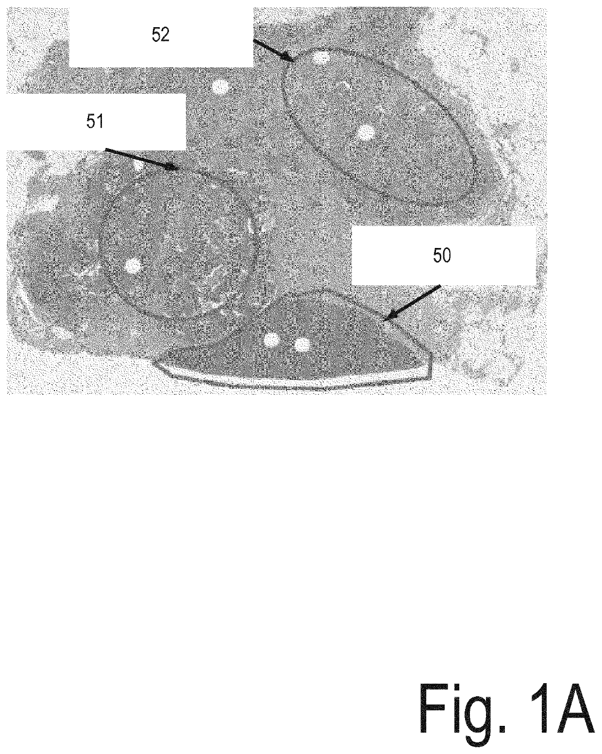
[0060]The data processing system 41 is configured to read microscopic image data generated using an image acquisition unit 45. In the exemplary embodiment, the image acquisition unit 45 is a microscope slide scanner, such as a whole slide scanner, which is configured to acquire an image of biologica...


PUM


Abstract

Disclosed is a system for analysis of microscopic image data acquired from biological cells. The system includes a data processing system which is configured to read the image data and determine a plurality of vertices, wherein each of the vertices represents a location of an entity of interest within a region of interest of the image data. The data processing system generates a plurality of graphs, wherein for each of the graphs, the generation of the respective graph includes generating a plurality of edges, wherein each of the edges has two of the plurality of vertices associated therewith. For each of the graphs one or more vertex sets are identified, each of which consisting of one or more of the plurality of vertices. The data processing system further determines, for each of the graphs, a number of the identified vertex sets.

Description

FIELD OF THE INVENTION[0001]The present invention relates to computational analysis of microscopic image data acquired from biological cells.BACKGROUND OF THE INVENTION[0002]Tissue pathology is a cornerstone in cancer diagnosis and prognosis. In conventional techniques of cancer diagnosis and prognosis, pathologists visually review stained slides of cancer biopsy samples and assign scores to the detected tumors. This process, however, is time-consuming and the results are often inconsistent across pathologists.[0003]Computer-assisted quantitative analysis of stained histology images have been made particularly efficient through whole slide scanners which allow acquisition of high resolution digital scans of entire microscopic slides. Such scanners can rapidly generate ultra-large 2D images of a whole tissue sample for digitization of histological slides. Automatic image processing procedures can then be applied to extract structures of interest from the original image for use in dia...

Claims


Application Information

Patent Timeline
Patent Type & Authority Applications(United States)
IPC IPC(8): G06T7/00G06K9/32G06K9/62
CPCG06T7/0012G06K9/3241G06T2207/20072G06T2207/30024G06K9/6273G06T2207/20084G06F18/2414
Inventor CHATTERJEA, SUPRIYOKORST, JOHANNES HENRICUS MARIAVAN LEEUWEN, MARINUS BASTIAANWIMBERGER-FRIEDL, REINHOLD
Owner KONINKLJIJKE PHILIPS NV
Features
  • R&D
  • Intellectual Property
  • Life Sciences
  • Materials
  • Tech Scout
  • Unparalleled Data Quality
  • Higher Quality Content
  • 60% Fewer Hallucinations
Learn More