

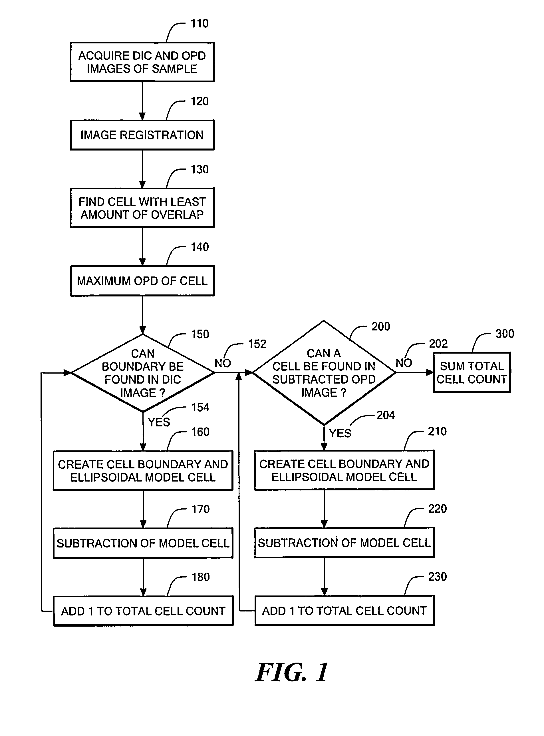
Phase subtraction cell counting method
a cell counting and phase subtraction technology, applied in the field of phase subtraction cell counting method, can solve the problems of inability to accurately count cells in cell clusters, slow change toward reducing the standard, low success rate, etc., and achieve the effect of accurately counting the number of cells and sufficient time for images
- Summary
- Abstract
- Description
- Claims
- Application Information
AI Technical Summary
Benefits of technology
Problems solved by technology
Method used
Image

Examples
example 1
Adapting a Microscope to Perform Optical Quadrature, DIC, and Epi-Fluorescence
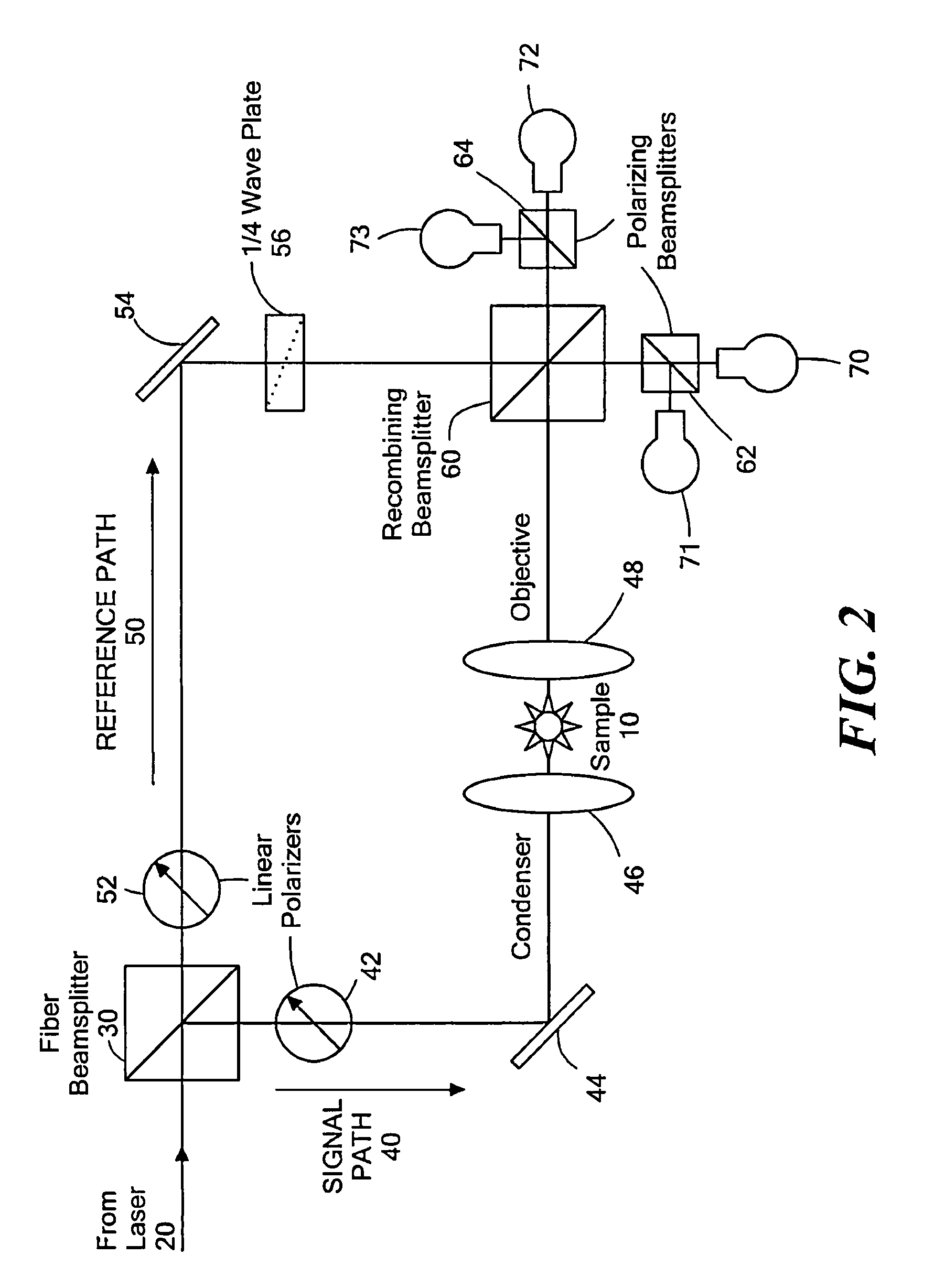
[0183]A Nikon ECLIPSE TE200 has one camera port on the base of the microscope, so all imaging modalities must be captured through a single output. The optics within the microscope are designed to produce the image of the sample at the end of the threads on the port because that is the location of the CCD chip if a CCD camera is attached. This configuration is ideal for capturing epi-fluorescence images because it contains the least amount of optics in the path that can reflect the fluorescent light. However, optical quadrature requires mixing of the reference path with the signal path, which cannot be done within the microscope base without major changes to the microscope body. Consequently, all CCD cameras were positioned away from the microscope body.
[0184]In the initial design, the microscope was already adapted for optical quadrature. An adjustable relay lens sent all light from the microscope to the r...
example 2
Cell Counting of Mouse Embryos Using DIC and Optical Quadrature Microscopy
[0191]Live mouse embryos at the 8-26 cell stage were counted using DIC and optical quadrature microscopic images according to the method of the present invention.
[0192]C57BL / 6J and CBA / CaJ strains of mice were purchased from the Jackson Laboratory (Bar Harbor, Me.) and bred in the laboratory. The mice were handled according to the Institutional Animal Care and Use Committee (IACUC) regulations in an Association for the Assessment and Accreditation of Laboratory Animal Care (AAALAC) approved facility. C57BL / 6 female mice, 2-6 months old, were superovulated according to standard procedures (Nagy et al., 2003). A first injection of 5 IU of eCG (Sigma, St. Louis, Mo.) was administered, followed 48 h later by 10 IU of hCG (Sigma). Individual C57BL / 6 female mice were mated overnight with a single CBA / Ca male and evaluated for the presence of a vaginal plug the next morning, indicating that mating had occurred. Two-c...
example 3
Software for Controlling the DIC / Fluorescence CCD
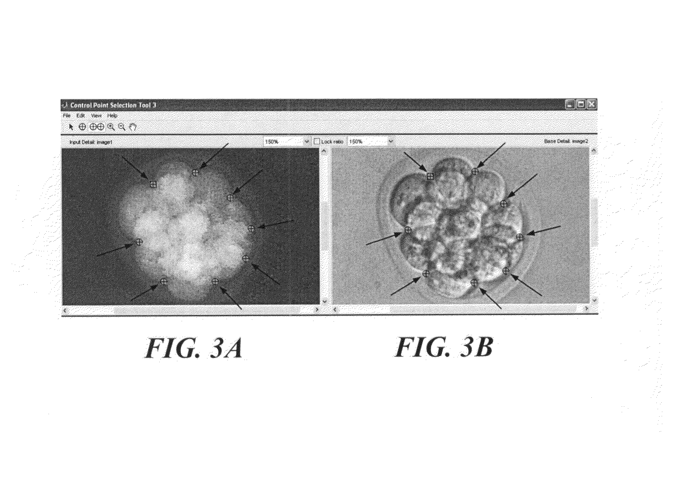
[0205]This Example contains software that controls the DIC / Fluorescence CCD. The list of files includes: dic_fl_main.m to provide the options to the user, dic_image.m to acquire DIC images, fl_image.m to acquire Epi-Fluorescence images, brightfield_image.m to acquire images that are not registered to the optical quadrature CCDs, fregister.m to produce a transform that registers the DIC / fluorescence CCD to the optical quadrature CCDs, registration.txt to display the commands required for fregister, and create_folder.m to produce the automatic folder structure. The acquired DIC and epi-fluorescence images are automatically registered to the optical quadrature CCDs by the last saved transform created by fregister. The text file, registration, contains the required commands to be entered on the Matlab program running on the computer controlling the optical quadrature CCDs to capture an image with camera 0 to register the DIC / fluorescence ...
PUM
 Abstract
Description
Claims
Application Information