Medical image conversion

a technology for medical images and conversion functions, applied in image data processing, medical data mining, radiation therapy, etc., can solve the problems of risk of introducing false information, and less accurate generated conversion functions, so as to minimize the penalty of said first and second penalties and optimize the conversion function accurately.

Inactive Publication Date: 2021-07-01
RAYSEARCH LAB
View PDF0 Cites 3 Cited by
  • Summary
  • Abstract
  • Description
  • Claims
  • Application Information

AI Technical Summary

Benefits of technology

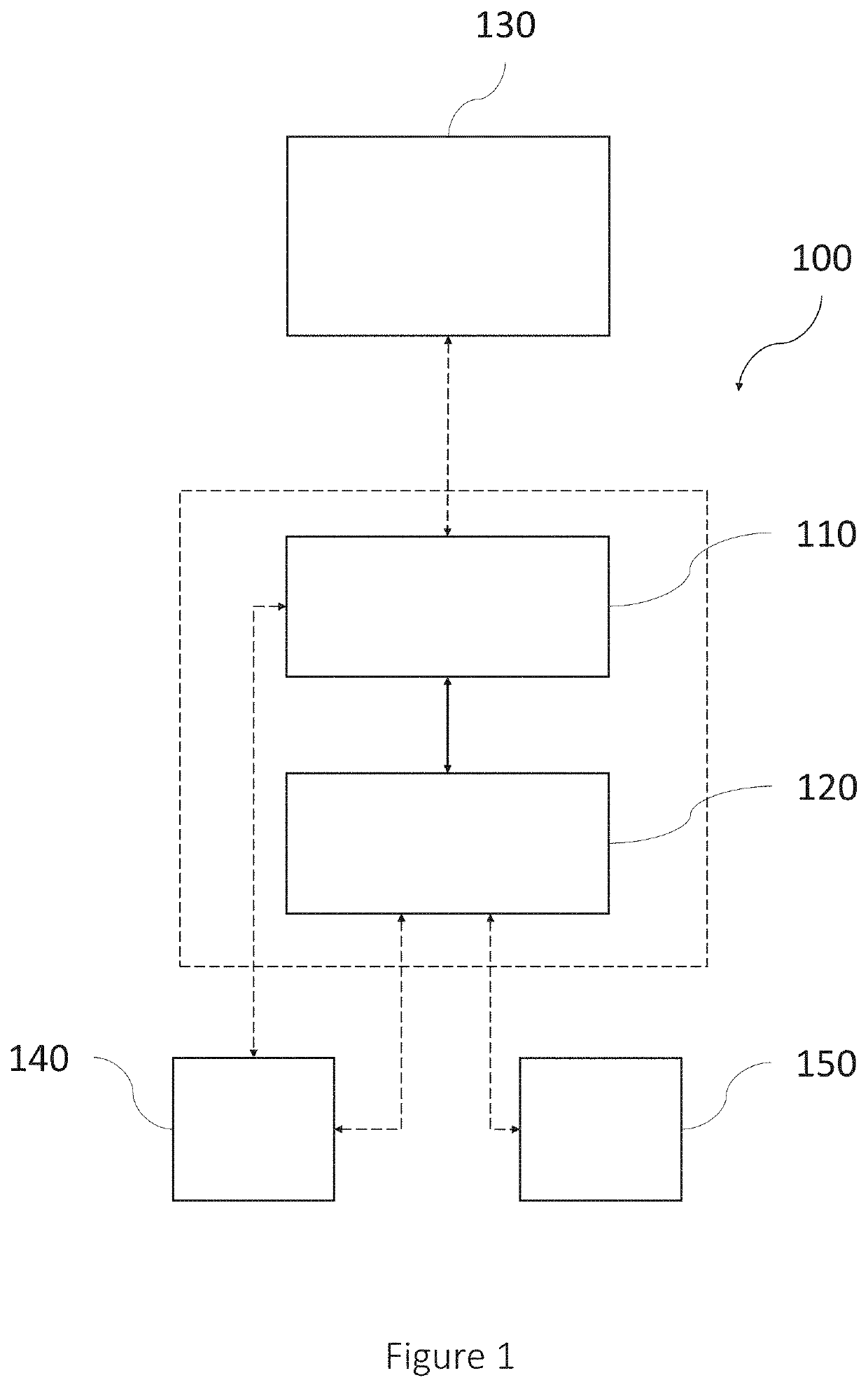
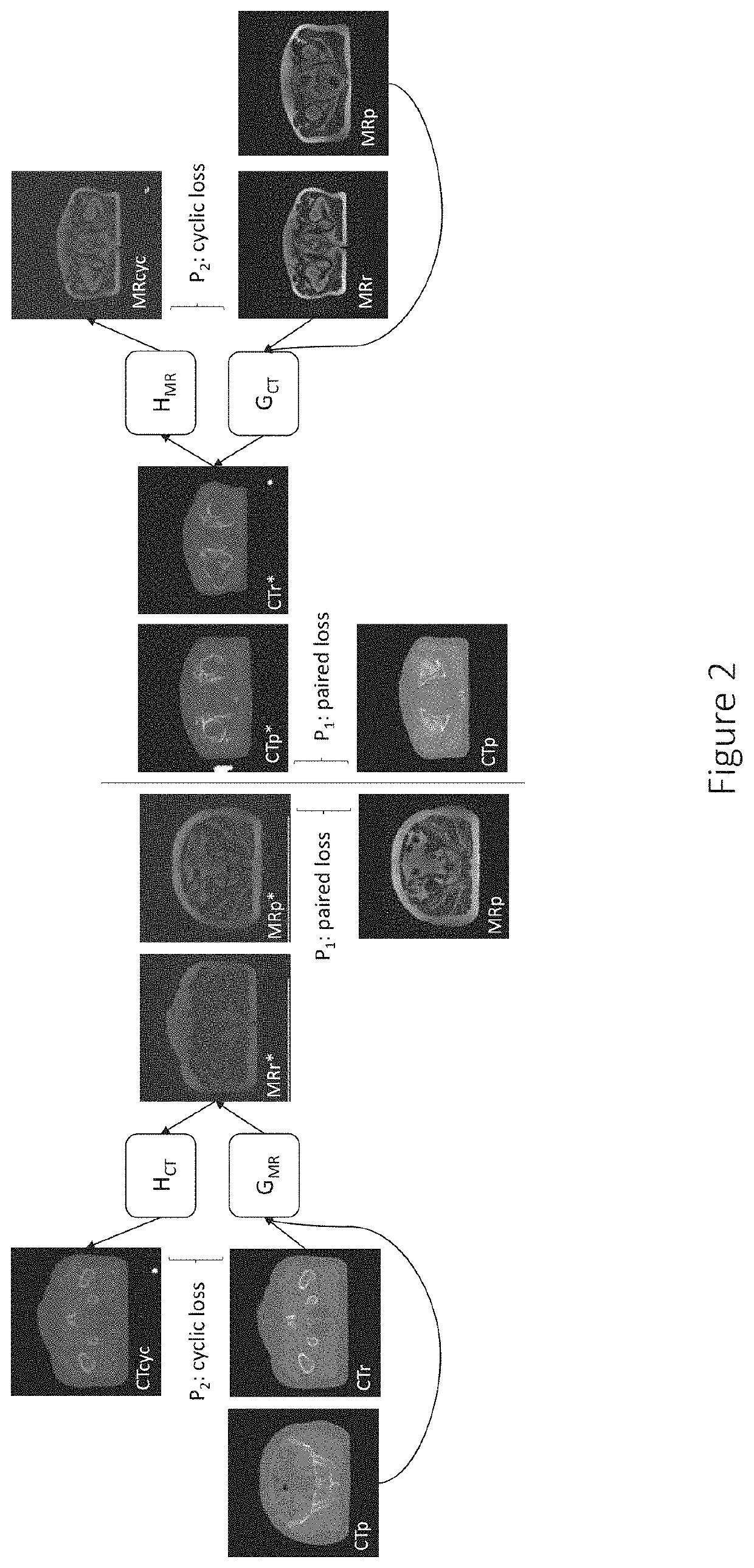
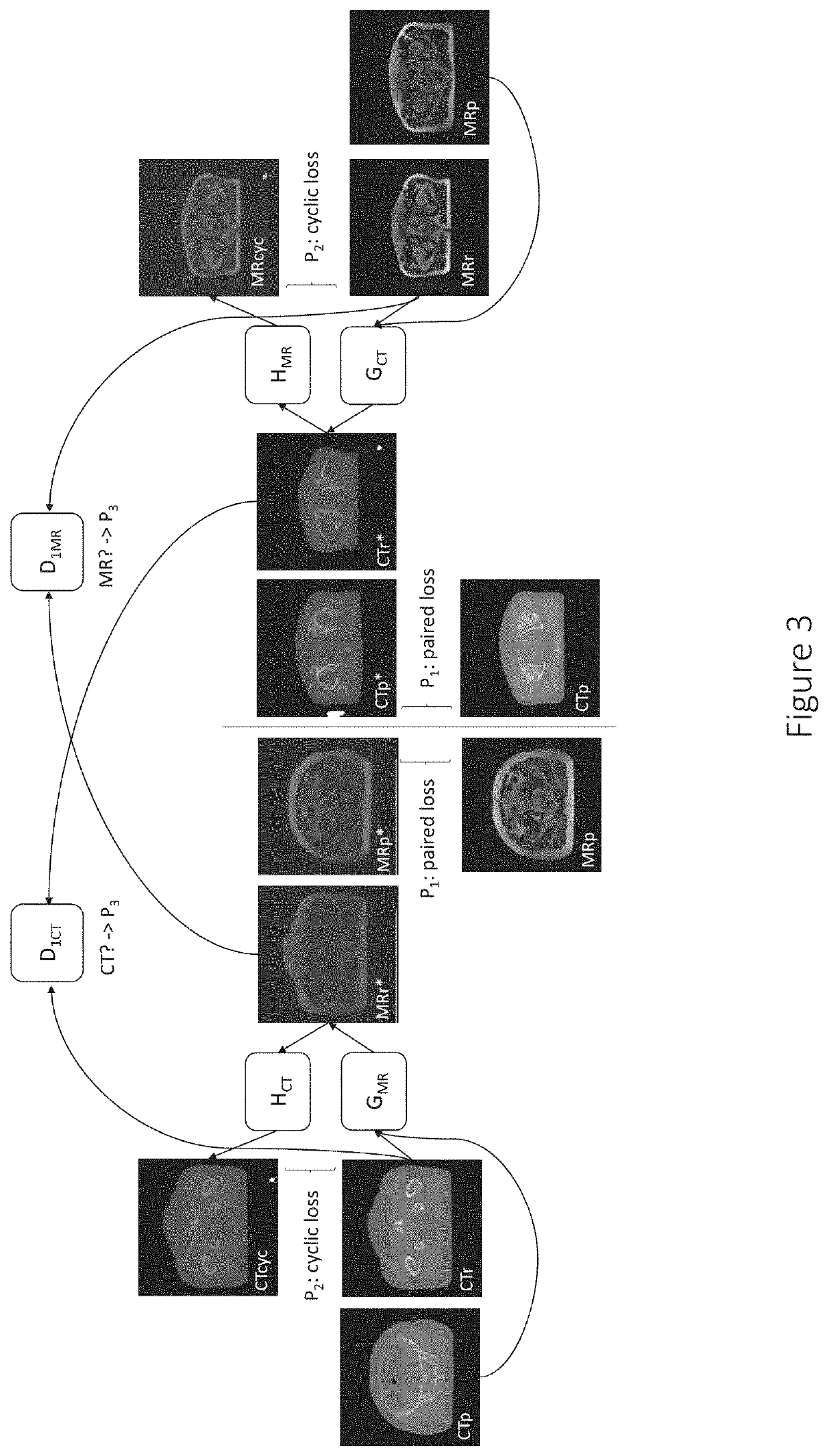
[0009]The above described problem is addressed by the claimed system for generating an optimized parametrized conversion function T for converting an original medical image of a first image type into a converted medical image of a second image type. The system may comprise at least one processing unit configured to: obtain original medical images of the first and second image types; obtain an initial parametrized conversion function G to convert original medical images of the first image type into converted medical images of the second image type; calculate a first penalty P1 based on at least one comparison of a first original medical image of the second image type with a first converted medical image of the second image type, that has been generated by applying a first parametrized conversion function G1, which is based on the initial parametrized conversion function G, to a first original medical image of the first image type that forms an image pair with the first original medical image of the second image type and has thereby been...

Problems solved by technology

In the approach described in the article Deep MR to CT Synthesis using Unpaired Data, the fact that only unpaired data is used may make the generated conversion function less accurate.
In this approach there is also a risk of introducing false information, which may nev...

Method used


Image

  • Medical image conversion
  • Medical image conversion
  • Medical image conversion

Examples


use case embodiment

[0092]To set the presently disclosed systems and methods in a larger context, the generating of an optimized parametrized conversion function T may in use case embodiments be preceded by the generating of original medical images.

[0093]FIG. 7 is a flow diagram exemplifying such a larger context, including the steps of: obtaining original medical images; obtaining an initial parametrized conversion function; calculating a first penalty P1 based on paired images, by comparing the converted medical image of the second image type with the paired original medical image of the second image type; calculating a second penalty P2 based on comparisons between converted medical images and original medical images, after converting them into images of the same image type; and generating an optimized parametrized conversion function based on the parameters of the initial parametrized conversion function and at least said first and second penalties P1 and P2. FIG. 7 further includes steps of: gener...


PUM


Abstract

In accordance with one or more embodiments herein, a system for generating an optimized parametrized conversion function T for converting an original medical image of a first image type into a converted medical image of a second image type is provided. The system comprises at least one processing unit configured to: obtain original medical images of the first and second image types; obtain an initial parametrized conversion function G to convert original medical images of the first image type into converted medical images of the second image type; calculate a first penalty P1 based on at least one comparison of a first original medical image of the second image type with a first converted medical image of the second image type, that has been generated by applying a first parametrized conversion function G1, which is based on the initial parametrized conversion function G, to a first original medical image of the first image type that forms an image pair with the first original medical image of the second image type and has thereby been determined to show the same part of the same patient; calculate a second penalty P2 based on at least one comparison of an original medical image of the first image type and a converted medical image of the second image type that has been generated by applying a second parametrized conversion function G2, which is based on the initial parametrized conversion function G, to the original medical image of the first image type, after converting the original medical image of the first image type and/or the converted medical image of the second image type into images of the same image type; and generate the optimized parametrized conversion function T based on the parameters of the initial parametrized conversion function G and at least said first and second penalties P1 and P2.

Description

TECHNICAL FIELD[0001]The present disclosure relates generally to systems and methods for generating an optimized parametrized conversion function T for converting an original medical image of a first image type into a converted medical image of a second image type.BACKGROUND[0002]Computed tomography (CT) imaging is useful for many purposes. CT images may e.g. be used for dose calculation in radiation treatment planning. However, there are reasons for avoiding CT imaging if other ways of obtaining images that can be used for dose calculation can be found—CT examination takes time, and patients are exposed to radiation.[0003]It is possible to use cone beam computed tomography (CBCT) images for dose calculation, but CBCT images are more prone to artefacts than CT images, and the intensity scale may also be different than in CT images.[0004]The article Deep MR to CT Synthesis using Unpaired Data (Wolterink et al. 2017) describes the training of a general adversarial network (GAN) to con...

Claims


Application Information

Patent Timeline
IPC IPC(8): G06T11/60G06T7/00G06T7/11G06T7/143G06T7/174
CPCG06T11/60G06T7/0014G06T7/11G06T7/143G06T2207/10088G06T2207/20084G06T2207/20081G06T2207/10081G06T7/174G01R33/5608G06T11/008A61N5/103G16H30/40G16H50/70G06T2211/464
Inventor ANDERSSON, SEBASTIANFREDRIKSSON, ALBINNORDSTRÖM, MARCUSNILSSON
Owner RAYSEARCH LAB
Why Eureka
Try Eureka