Ultrasensitive method for multiplexed detection of biomarkers

a biomarker and multiplexing technology, applied in the field of ultrasensitive methods for multiplexing detection of biomarkers, can solve the problems of imposing mental and physical burdens on subjects, unable to use multiplexing detection based on a single biomarker, and unable to accurately diagnose the level of only one biomarker

Pending Publication Date: 2021-07-08
SEOUL NAT UNIV R&DB FOUND
View PDF0 Cites 2 Cited by
  • Summary
  • Abstract
  • Description
  • Claims
  • Application Information

AI Technical Summary

Problems solved by technology

Since the levels of several biomarkers for one disease vary simultaneously or the level of one biomarker varies by several diseases, the level of only one biomarker cannot be used for an accurate diagnosis.
Since ELISA-based techniques cannot be used for multiplexed detection, the detection of a number of biomarkers requires a sufficient amount of blood and a sufficient number of detection kits in proportion to the number of the biomarkers.
The sampling of a large amount of blood imposes mental and physical burdens on

Method used


Image

  • Ultrasensitive method for multiplexed detection of biomarkers
  • Ultrasensitive method for multiplexed detection of biomarkers
  • Ultrasensitive method for multiplexed detection of biomarkers

Examples


example 1

ization of FRET-PAINT

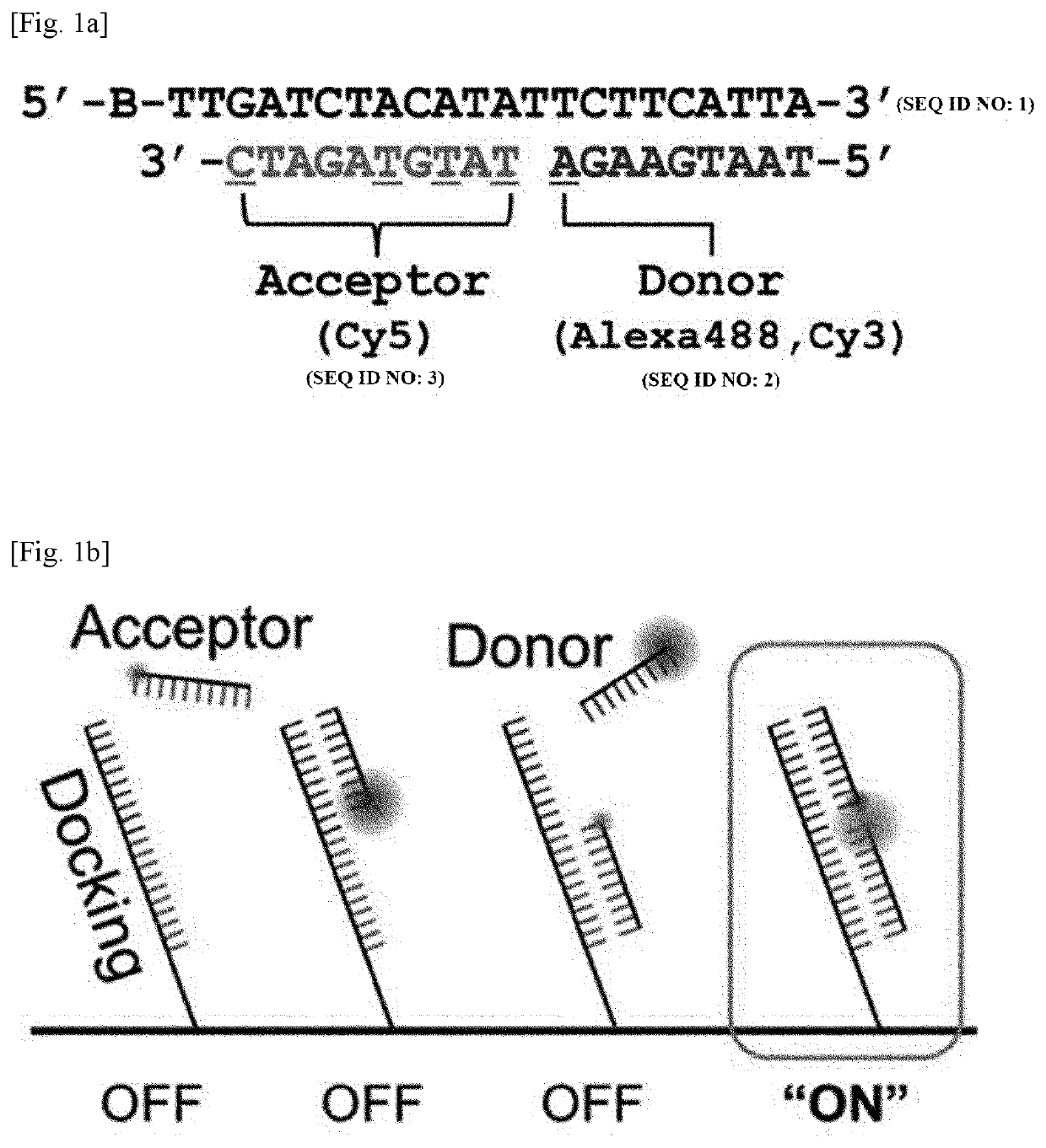
[0114]Surface-immobilized DNA strands and a TIRF microscope were used to test the feasibility of FRET-PAINT microscopy.

[0115]As shown in FIG. 1a, FRET-PAINT used three DNA strands (docking, donor, and acceptor strands). The docking strand (Docking_P0) labeled with a biotin at the 5′-end has two docking sites, each of which base-pairs with the donor or acceptor strand. To increase the FRET probability, a shorter length for the donor strand than for the acceptor strand was chosen whereas relatively longer acceptor strand was used. The donor strand was labeled at the 3′-end with Alexa488 (Donor_P1_Alexa488) whereas the acceptor strand was labeled with Cy5 (Acceptor_P11_Cy5) at the 3′-end.

[0116]The sequences of the strands are as follows:

(SEQ ID NO: 1)Docking_PO = 5′-Biotin-TTGATCTACATATTCTTCATTA-3′(SEQ ID NO: 2)Donor_P1_Cy3 - 5′-TAATGAAGA-Cy3-3′(SEQ ID NO: 2)Donor_P1_Alexa488 = 5′-TAATGAAGA-Alexa488-3′(SEQ ID NO: 3)Acceptor_P2_Cy5 = 5′-Cy5-TATGTAGATC-3′(SEQ ID NO: ...

example 2

lution Imaging with DNA-PAINT and FRET-PAINT

[0124]The following experimental procedure was performed to compare the super-resolution imaging speeds of DNA-PAINT and FRET-PAINT.

[0125]First, microtubules of COS-7 cells were immunostained with the anti-tubulin antibody which is labeled with Docking_P1.

[0126]Then, for DNA-PAINT, microtubules were imaged after injecting 1 nM Cy5-labeled imager strand (Acceptor_P2′_Cy5). For FRET-PAINT, microtubules of the same area were imaged after injecting 30 nM donor (Donor_P1_Alexa488) and 20 nM acceptor (Acceptor_P2_Cy5) strands. Single-molecule images were recorded at a frame rate of 10 Hz, which is fast enough to reliably detect binding of donor and acceptor strands.

[0127]To quantitatively compare the imaging speed of DNA-PAINT and FRET-PAINT, the number of spots of FIGS. 2a and 2b was measured. The same analysis was performed for nine additional imaging areas and the averaged speeds were compared. The convolved resolutions computed as ((localiza...

example 3

T Multiplexed Imaging

[0130]The multiplexing capability of FRET-PAINT microscopy was assessed by the following experimental procedure.

[0131]Microtubules and mitochondria of COS-7 cells were immunostained using anti-tubulin antibody and anti-Tom20 antibody, respectively. The anti-tubulin antibody and anti-Tom20 antibody were orthogonally conjugated with Docking_P1 and Docking_P2, respectively. In the present invention, two approaches were used for multiplexed imaging.

[0132]The results are shown in FIGS. 3a to 3h. In the one approach shown in FIG. 3a (by sequential treatment with donor and acceptor strands), microtubules were first imaged by injecting 20 nM Donor_P1_Alexa488 and 10 nM Acceptor_P2_Cy5 (FIG. 3b) and mitochondria were then imaged by injecting 10 nM Donor_P2_Alexa488 and 10 nM Acceptor_P2_Cy5 (FIGS. 3c and 3d). In the second approach shown in FIG. 3e (by simultaneous treatment with donor and acceptor strands), after injection of all DNA probes (10 nM Donor_P1_Cy3 for micro...


PUM

PropertyMeasurementUnit
Ratioaaaaaaaaaa
Wavelengthaaaaaaaaaa
Fluorescenceaaaaaaaaaa

Abstract

The present invention relates to an ultrasensitive method for multiplexed detection of biomarkers. More specifically, the present invention provides a method for multiplexed detection of biomarkers, including a) attaching one or more types of biomarkers present in a sample taken from a subject to the surface of a substrate, b) attaching docking strands to the biomarkers and allowing detection antibodies to specifically bind to the biomarkers, and binding the detection antibodies to the corresponding biomarkers, c) binding imager strands labeled with fluorescent molecules or combinations of donor and acceptor strands to the docking strands to generate a fluorescence signal, and d) detecting the fluorescence signal. Steps c) and d) are repeated as many times as the number of the biomarker types by removing the used imager strands or the used donor and acceptor strands and introducing new imager strands or different combinations of donor and acceptor strands to the docking strands.

Description

TECHNICAL FIELD[0001]This application claims priority to Korean Patent Application No. 10-2018-0029473 filed on Mar. 13, 2018, the disclosure of which is incorporated herein by reference in its entirety.[0002]The present invention relates to an ultrasensitive method for multiplexed detection of biomarkers. More specifically, the present invention relates to a method for multiplexed detection of biomarkers, including a) attaching one or more types of biomarkers present in a sample taken from a subject to the surface of a substrate, b) attaching docking strands to the detection antibodies and allowing detection antibodies to specifically bind to the corresponding biomarkers, c) binding imager strands labeled with fluorescent molecules or combinations of donor and acceptor strands to the docking strands to generate a fluorescence signal, and d) detecting the fluorescence signal, wherein steps c) and d) are repeated as many times as the number of the biomarker types by removing the used...

Claims


Application Information

Patent Timeline
IPC IPC(8): G01N33/543C12Q1/6834C12Q1/6818G01N21/64
CPCG01N33/54306C12Q1/6834G01N2021/6439G01N21/6428C12Q1/6818G01N2458/10G01N33/542G01N21/6408G01N33/543G01N33/582C12Q1/6804C12Q2565/101C12Q2563/179C12Q2563/107
Inventor HOHNG, SUNGCHULLEE, JONGJIN
Owner SEOUL NAT UNIV R&DB FOUND
Why Eureka
Try Eureka