
Microscopy method and microscope for producing an image of an object

a technology of electronic image and microscope, which is applied in the field of microscopy method and microscope for producing electronic image of objects, can solve the problems of not providing additional image information through digital magnification, and no increase in light sensitivity achieved by digital zooming, so as to improve the image of objects

Active Publication Date: 2021-07-29
CARL ZEISS MEDITEC AG
View PDF0 Cites 0 Cited by
  • Summary
  • Abstract
  • Description
  • Claims
  • Application Information

AI Technical Summary

Benefits of technology

The invention provides a microscopy method and microscope that can improve the image of an object by reducing the optical imaging scale. By doing so, the intensity of the radiation from the object is increased on fewer pixels of the image detector, resulting in a higher signal-to-noise ratio. Additionally, the depth of field range of the image is increased, improving the overall image quality. The method and microscope can automatically optimize these parameters without user intervention, providing a better image without compromising the resolution. The microscope can also adjust the working distance and the illuminance on the object to further improve the image quality.

Problems solved by technology

However, all image information is contained in the optical image and digital magnification does not provide additional image information.
In particular, no increased light sensitivity is achieved by digital zooming.

Method used


Image

  • Microscopy method and microscope for producing an image of an object
  • Microscopy method and microscope for producing an image of an object
  • Microscopy method and microscope for producing an image of an object

Examples


Embodiment Construction

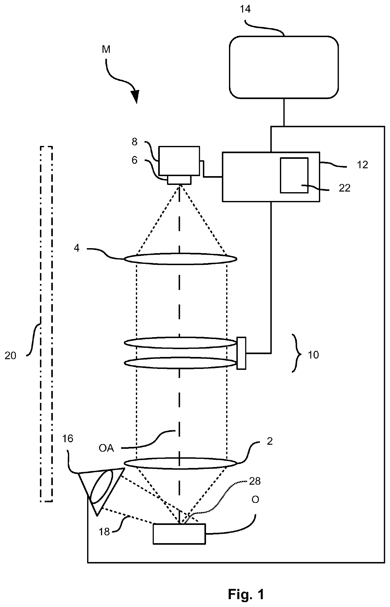
[0048]FIG. 1 schematically shows a microscope M for imaging an object O. Preferably, the microscope M is a surgical microscope which images part of a patient during a surgical intervention. The microscope M images the object O from an object field on an image field, lying on a detector area 6 of an image detector 8, via an objective 2 and a tube lens 4. A zoom optical unit 10 is present in the beam path, the zoom optical unit altering the optical imaging scale, for example, the size of the image field in the case of an unchanging size of the object field, and including to this end a drive not denoted in any more detail. The optical imaging scale, with which the object field is imaged on the image field by the objective 2, the zoom optical unit 10 and the tube lens 4, is therefore adjustable. The image detector 8 is read by a controller 12, which converts the data obtained in this way into an electronic image, which is displayed on a display device 14.

[0049]The object O is illuminate...


PUM


Abstract

A microscopy method is for producing an electronic image of an object, wherein the object is imaged with an adjustable optical imaging scale on an image detector. The method includes: selecting a parameter for the electronic image, wherein the parameter can be influenced by the optical imaging scale and differs from the image field dimensions, and setting a setpoint value range for the parameter, setting a total imaging scale for the electronic image, wherein adjusting or controlling the parameter of the electronic image is implemented such that, at the same time, the parameter of the electronic image lies in the specified setpoint value range with a tolerance and the set total imaging scale is obtained, wherein the optical imaging scale forms a basis for a manipulated variable of the adjustment or closed-loop control and a digital image magnification is carried out on the basis of the set total imaging scale.

Description

CROSS REFERENCE TO RELATED APPLICATION[0001]This application claims priority of German patent application no. 10 2020 101 880.0, filed Jan. 27, 2020, the entire content of which is incorporated herein by reference.FIELD OF THE INVENTION[0002]The disclosure relates to a microscopy method and a microscope for producing an electronic image of an object, wherein the object is imaged with an adjustable optical imaging scale on an image detector.BACKGROUND OF THE INVENTION[0003]Surgical microscopes are known from US 2008 / 0266657 A1 or US 2005 / 0063047 A1. Further, digital surgical microscopes which include a digital magnification (often called zoom) combined with optical image magnification are known, by way of example, from US 2006 / 0092505 A1, US 2012 / 0002274 A1, U.S. Pat. No. 10,139,613 B2 or US 2008 / 0198448 A1.[0004]In microscopes which generate electronic images at adjustable optical magnification, usually the maximum optical magnification is utilized and additional zooming is performe...

Claims


Application Information

Patent Timeline
Patent Type & Authority Applications(United States)
IPC IPC(8): G02B21/36G02B21/06G02B21/00H04N5/232H04N5/225
CPCG02B21/365G02B21/06H04N5/2256H04N5/23296G02B21/0012H04N23/56H04N23/69
Inventor REGENSBURGER, ALOISNIETEN, CHRISTOPHHOEGELE, ARTURHAUGER, CHRISTOPH
Owner CARL ZEISS MEDITEC AG
Learn More