
Soluble Polypeptides and Methods of Using Same for Inhibiting Leukemia Inhibitory Factor Activity
- Summary
- Abstract
- Description
- Claims
- Application Information
AI Technical Summary
Benefits of technology
Problems solved by technology
Method used
Image

Examples
example 1
Engineered Soluble LIFR Polypeptides
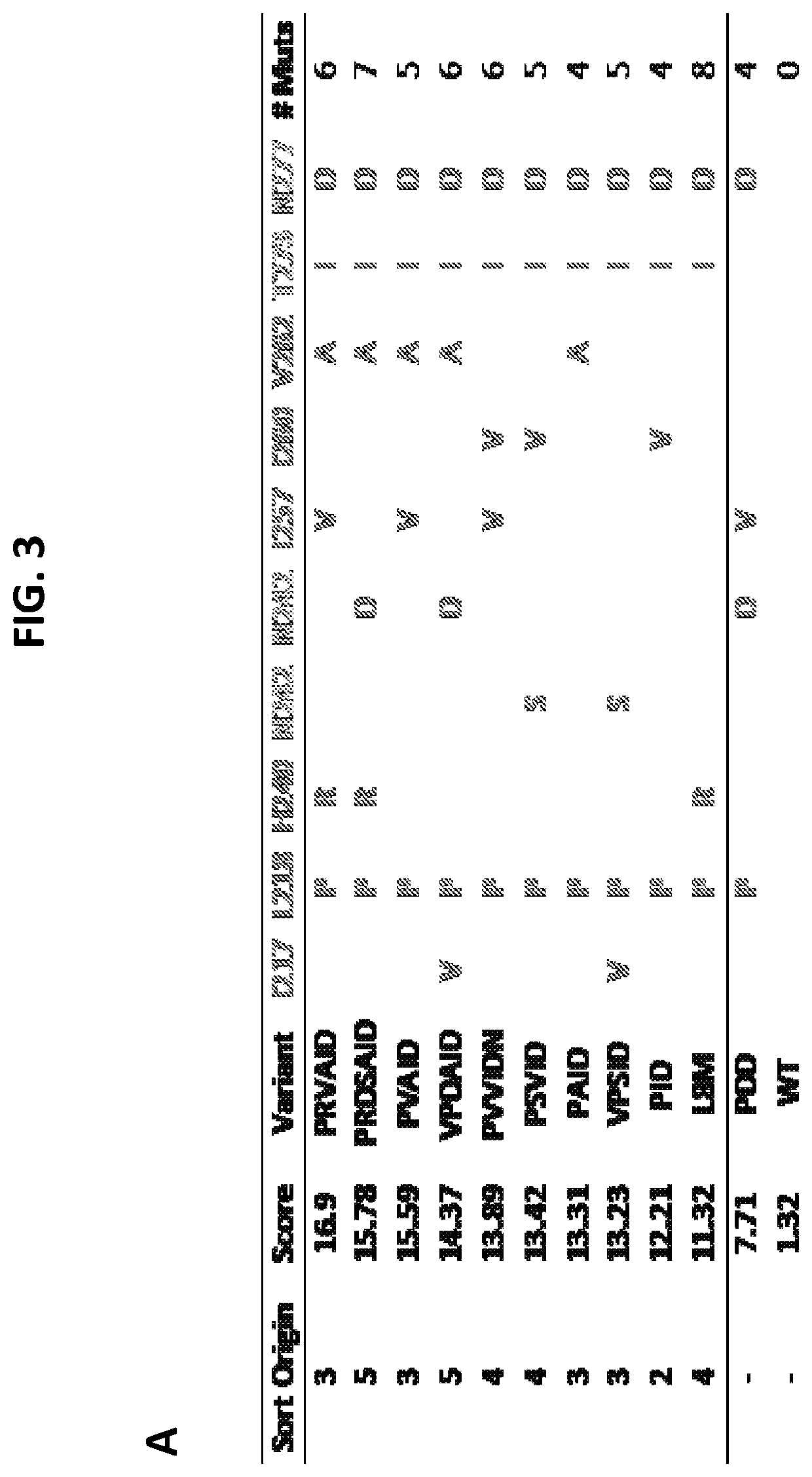
[0235]In this example, LIFR variants were identified from libraries of the Ig-like domain of human LIFR displayed on the surface of yeast and sorted using FACS for optimal binders to human LIF. After multiple rounds of sorting, variants were selected, sequenced, and characterized. LIFR variants isolated from the sorts are summarized in FIG. 3.
[0236]FIG. 3, panel A, shows consensus mutations observed in LIFR Ig-like domain variants isolated across multiple sorts. Numbering starts at the beginning of the LIFR Cytokine Binding Motif I (CBMI) domain. FIG. 3, panel B, shows binding scores of LIFR variants calculated from normalized, summed fluorescence binding values at low (10 pM, 100 pM) LIF concentrations. All scores were normalized to wild-type and for differential expression on the surface of yeast.
[0237]Based on the identified LIFR mutations (shown in FIG. 3, panel A), multiple combinations of possible mutant LIFR variants which were not observed...
example 2
Engineered Soluble GP130 Polypeptides
[0239]In this example, gp130 variants were identified from libraries of the CBM domain of human gp130 displayed on the surface of yeast and sorted using FACS for optimal binders to human LIF. After multiple rounds of sorting, variants were selected, sequenced, and characterized. gp130 variants isolated from the sorts are summarized in FIG. 6.
[0240]FIG. 6, panel A, shows consensus mutations observed in gp130 CBM domain variants isolated across multiple sorts. Numbering starts at the beginning of the gp130 Cytokine Binding Motif (CBM) domain. FIG. 6, panel B, shows a summary of mutant gp130 variants created using site-directed mutagenesis. FIG. 6, panel C, shows the binding scores of gp130 variants calculated as described in FIG. 3, panel B, using 100pM and 1nM as low LIF concentrations. Expression normalized to wild-type (WT) is also shown. From this analysis, ELDME (K45E-F46L-Y95D-I130M-K184E) was identified as the optimal gp130 variant.
[0241]In ...
example 3
Binding of LIFR and GP130 Variants to Human LIF
[0242]In this example, LIFR or gp130 variants were displayed on the surface of yeast. Binding to human LIF was measured via fluorescent antibody detection using flow cytometry.
[0243]FIG. 8, panel A, shows binding results of yeast displayed wild-type LIFR and LIFR variants to human LIF. Results for LIFR wild-type (WT) (circles), the “PDD” variant (L218P-N42D-N277D) (squares), and the VPRVVAID variant (mutations in FIG. 4, panel A) (triangles) are shown. The KD of the VPRVVAID variant was measured at 30 pM, which is 32-fold higher affinity compared to wild-type (WT).
[0244]FIG. 8, panel B, shows binding results of yeast displayed wild-type gp130 and gp130 variants to human LIF. Results for gp130 wild-type (WT) (circles), the “8M” variant (E4K-K5R-N14D-K45E-F46L-K83R-Y95D-N100S) (squares), and the ELDME variant (mutations in FIG. 4, panel A) (triangles) are shown. The KD of the ELDME variant was measured at 5 nM, which is 12-fold higher aff...
PUM
| Property | Measurement | Unit |
|---|---|---|
| Fraction | aaaaa | aaaaa |
| Fraction | aaaaa | aaaaa |
| Fraction | aaaaa | aaaaa |
Abstract
Description
Claims
Application Information