Assessment of Abnormality Regions Associated with a Disease from Chest CT Images

a computed tomography and abnormality assessment technology, applied in image enhancement, instruments, applications, etc., can solve the problems of false negatives, limited availability of rt-pcr test kits, and the death of covid-19 to a significant percentage of infected patients

Active Publication Date: 2021-09-30
SIEMENS MEDICAL SOLUTIONS USA INC
View PDF6 Cites 1 Cited by
  • Summary
  • Abstract
  • Description
  • Claims
  • Application Information

AI Technical Summary

Benefits of technology

[0005]In accordance with one or more embodiments, systems and methods for assessing a disease are provided. Medical imaging data of lungs of a patient is received. The lungs are segmented from the medical imaging data and abnormality regions associated with a disease are segmented from the medical imaging data. An assessment of the disease is determined based on the segmented lungs and the segmented abnormality regions. In one embodiment, the disease may be COVID-19 (coronavirus disease 2019) and the abnormality regions associated with COVID-19 comprise opacities of one or more of ground glass opacities (GGO), consolidation, and crazy-paving pattern. However, the disease may be any other disease, such as, e.g., SARS (severe acute respiratory syndrome), MERS (Middle East respiratory syndrome), other types of viral pneumonia, bacterial pneumonia, fungal pneumonia, mycoplasma pneumonia, and other types of pneumonia.

Problems solved by technology

However, COVID-19 is fatal to a significant percentage of infected patients.
However, the sensitivity of RT-PCR has been found to be as low as 60 to 70%, potentially resulting in false negatives.
Additionally, limited availability of RT-PCR test kits has contributed to the undetected spread of COVID-19.

Method used


Image

  • Assessment of Abnormality Regions Associated with a Disease from Chest CT Images
  • Assessment of Abnormality Regions Associated with a Disease from Chest CT Images
  • Assessment of Abnormality Regions Associated with a Disease from Chest CT Images

Examples


Embodiment Construction

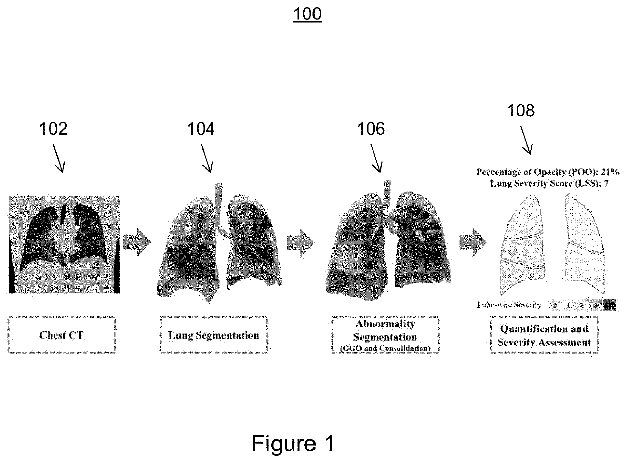
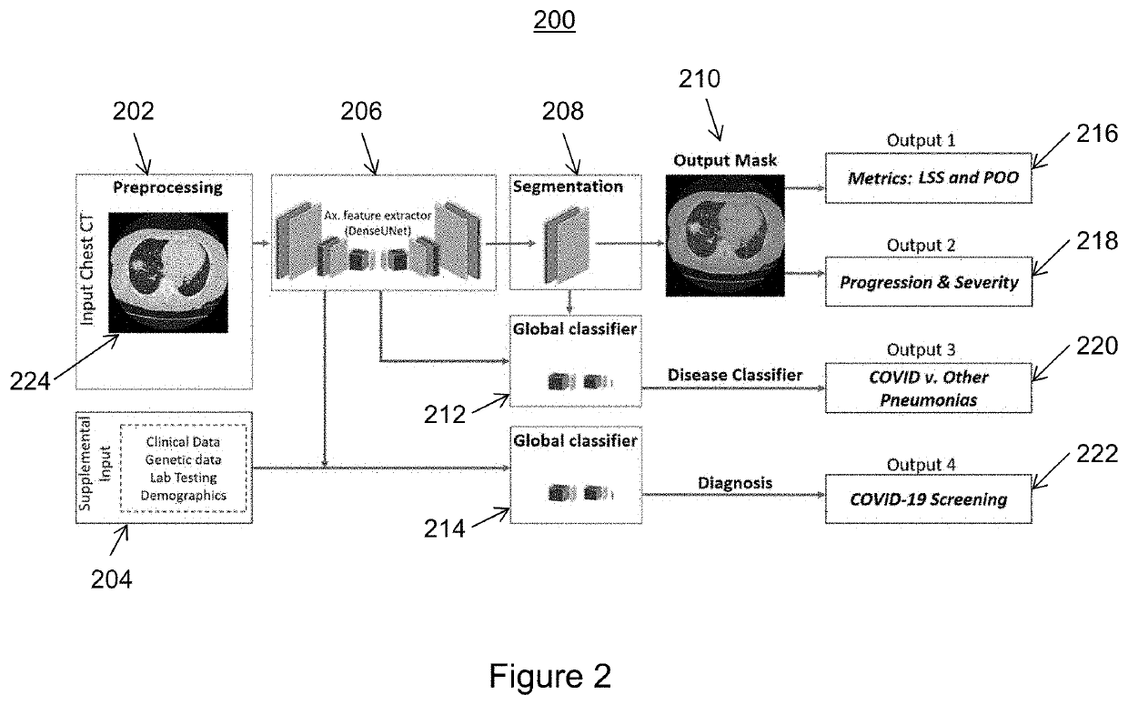
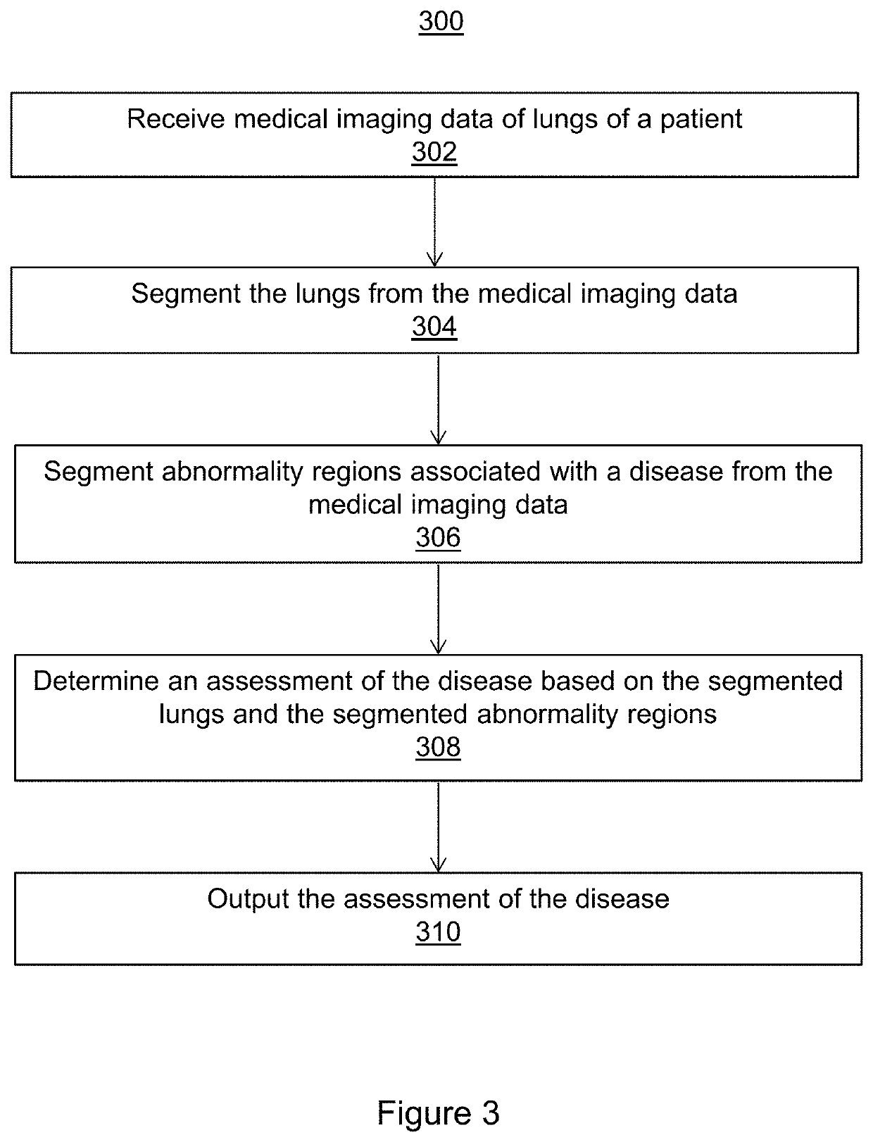
[0019]The present invention generally relates to methods and systems for the assessment of abnormality regions associated with COVID-19 (coronavirus disease 2019) from chest CT (computed tomography) images. Embodiments of the present invention are described herein to give a visual understanding of such methods and systems. A digital image is often composed of digital representations of one or more objects (or shapes). The digital representation of an object is often described herein in terms of identifying and manipulating the objects. Such manipulations are virtual manipulations accomplished in the memory or other circuitry / hardware of a computer system. Accordingly, is to be understood that embodiments of the present invention may be performed within a computer system using data stored within the computer system.

[0020]COVID-19 is an infectious disease that typically presents such respiratory symptoms as fever, cough, and difficulty breathing. CT imaging of the lungs of patients th...


PUM


Abstract

Systems and methods for assessing a disease are provided. Medical imaging data of lungs of a patient is received. The lungs are segmented from the medical imaging data and abnormality regions associated with a disease are segmented from the medical imaging data. An assessment of the disease is determined based on the segmented lungs and the segmented abnormality regions. The disease may be COVID-19 (coronavirus disease 2019) or diseases, such as, e.g., SARS (severe acute respiratory syndrome), MERS (Middle East respiratory syndrome), or other types of viral and non-viral pneumonia.

Description

CROSS-REFERENCE TO RELATED APPLICATIONS[0001]This application claims the benefit of U.S. Provisional Application No. 63 / 002,457, filed Mar. 31, 2020, the disclosure of which is incorporated herein by reference in its entirety.TECHNICAL FIELD[0002]The present invention relates generally to the assessment of abnormality regions associated with a disease from chest computed tomography images, and in particular to the assessment of abnormality regions associated with COVID-19 (coronavirus disease 2019).BACKGROUND[0003]COVID-19 (coronavirus disease 2019) is an infectious disease caused by the severe-acute respiratory symptom coronavirus 2 (SARS-Cov2). Common symptoms of COVID-19 include fever, cough, and difficulty breathing. In severe cases, COVID-19 can cause pneumonia, severe acute respiratory syndrome, and multiple organ failure. In the majority of cases, patients infected with COVID-19 experience mild to moderate symptoms that do not require hospitalization. However, COVID-19 is fat...

Claims


Application Information

Patent Timeline
Patent Type & Authority Applications(United States)
IPC IPC(8): G06T7/00G06T7/11G06T11/00
CPCG06T7/0016G06T7/11A61B5/055G06T2207/10081G06T2207/30061G06T11/008G06T7/0012G06T7/62G06N3/08G06T2207/20081G06T2207/20084G06N3/045G06F18/214G06F18/24A61B6/5217A61B8/5223
Inventor CHAGANTI, SHIKHAGRBIC, SASAGEORGESCU, BOGDANXU, ZHOUBINGLIU, SIQIYOO, YOUNGJINRE, THOMASCHABIN, GUILLAUMEFLOHR, THOMASZIEBANDT, VALENTINCOMANICIU, DORINTEIXEIRA, BRIANPIAT, SEBASTIEN
Owner SIEMENS MEDICAL SOLUTIONS USA INC
Why Eureka
Try Eureka