3D analysis with optical coherence tomography images

a technology of optical coherence tomography and 3d analysis, applied in image analysis, image enhancement, medical science, etc., can solve the problems of lack of depth resolution of density measurement in 2d projection images, various deficiencies and limitations, and artifacts in vessel segmentation

Pending Publication Date: 2021-10-14
KK TOPCON
View PDF7 Cites 0 Cited by
  • Summary
  • Abstract
  • Description
  • Claims
  • Application Information

AI Technical Summary

Benefits of technology

The present patent describes a method for quantifying the three-dimensional structure of an object using optical coherence tomography (OCT) data. The method involves pre-processing the OCT data, segmenting the data to identify specific parts of the object, and analyzing the data to determine its size and shape. This information can then be visualized using a two-dimensional map. The method can be applied to a variety of objects, such as the retina and choroidal vasculature, and can help to improve the accuracy and reliability of OCT data analysis.

Problems solved by technology

However, due to inherent properties of OCT imaging, artifacts in vessel segmentation will emerge if the thresholding is directly applied to the images.
Other techniques have thus been developed to segment components of OCT data, but these too suffer from various deficiencies and limitations.
Using a choroidal vessel density measurement in 2D projection images lacks depth resolution and can suffer from shadow artifact.
Similarly, automated detection of vessel boundaries (even with machine-learning) can be affected by shadow artifacts, and is additionally limited to application in two-dimensional (2D) B-scans only and for larger vessels.
Further, the segmented vessel continuity may be poor due the segmentation is repeated for each B-scan in a volume, rather than applied to the volume as a whole.
Other segmentation techniques are only applicable for normal (non-diseased eyes) and suffer errors when retinal structure changes due to diseases.
Further, some segmentations are subject to inaccuracies related to the application of noise reduction filters on underlying data.
As a result, the segmentation can be limited in dimension and location.
Because of these limitations it has not been practical and / or not even possible to present many clinically valuable visualizations and quantifications of choroidal vasculature.
This greatly diminishes the value of, and does not fully utilize, the data.
In other instances, the quantifications are taken from OCT data that remains too noisy to perform an accurate analysis, utilize averages taken from many volumes, which can still suffer from noise and also requires increased scanning times (for each iterative volume form which the average is taken), or are limited to relatively small regions of interest (e.g., 1.5 mm under the fovea in single B-scan).
Accordingly, medical practitioners have not been able to fully appreciate clinically pertinent information available 3D volumetric OCT data.

Method used


Image

  • 3D analysis with optical coherence tomography images
  • 3D analysis with optical coherence tomography images
  • 3D analysis with optical coherence tomography images

Examples


Embodiment Construction

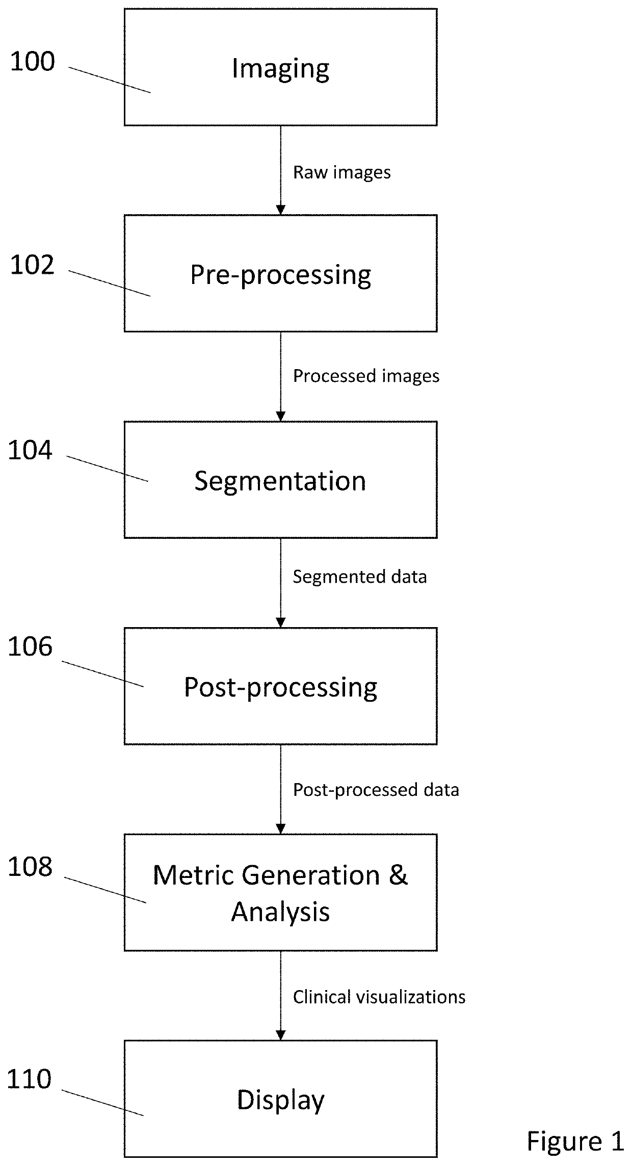
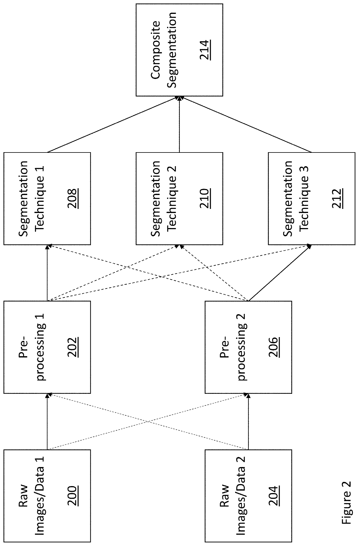
[0014]The present disclosure relates to clinically valuable analyses and visualizations of three-dimensional (3D) volumetric OCT data that was not previously practical and / or possible with known technologies. Such analyses and visualizations may improve a medical practitioner's ability to diagnose disease, monitor, and manage treatment. Briefly, the analysis is performed on, and the visualizations are created by, segmenting OCT data for a component of interest (e.g., choroidal vasculature) in three dimensions following a series of pre-processing techniques. The segmentation can be applied to the data following pre-processing, and then combined to produce a final full 3D segmentation of the desired component. Post-processing, such as a smoothing technique, may be then applied to the segmented component. While choroidal vasculature of OCT data is particularly discussed herein, the disclosure is not to be so limited.

[0015]An example method for producing clinically valuable analyses and...


PUM


Abstract

A method for generating clinically valuable analyses and visualizations of 3D volumetric OCT data by combining a plurality of segmentation techniques of common OCT data in three dimensions following pre-processing techniques. Prior to segmentation, the data may be subject to a plurality of separately applied pre-processing techniques.

Description

BACKGROUND OF THE INVENTION[0001]Optical coherence tomography (OCT) is a technique for in-vivo imaging and analysis of various biological tissues (as, for example, two-dimensional slices and / or three-dimensional volumes). Images created from three-dimensional (3D) volumetric OCT data show different appearances / brightness for different components of the imaged tissue. Based on this difference, those components can be segmented out from the images for further analysis and / or visualization. For example, choroidal vasculature has a darker appearance than choroidal stroma in OCT images. Therefore, the choroidal vasculature in OCT images can be segmented out by applying an intensity threshold. However, due to inherent properties of OCT imaging, artifacts in vessel segmentation will emerge if the thresholding is directly applied to the images. Other techniques have thus been developed to segment components of OCT data, but these too suffer from various deficiencies and limitations.[0002]Fo...

Claims


Application Information

Patent Timeline
Patent Type & Authority Applications(United States)
IPC IPC(8): G06T7/00G06T7/11G06T5/00A61B3/10
CPCG06T7/0012G06T7/11G06T5/002G06T2207/30101G06T2207/10101G06T2207/20108G06T2207/30041A61B3/102G06T7/62G06T5/70
Inventor MEI, SONGMAO, ZAIXINGSUI, XINWANG, ZHENGUOCHAN, KINPUI
Owner KK TOPCON
Why Eureka
Try Eureka