Systems and methods for analysis of tissue images

a tissue image and system technology, applied in the field of image processing, can solve the problems of difficult or impossible correlation to other nearby anatomies, and achieve the effects of reducing the rate of repeat procedures, reducing the rate of re-admission, and increasing the procedure yield ra

Active Publication Date: 2022-03-24
NUCLAI LTD
View PDF0 Cites 1 Cited by
  • Summary
  • Abstract
  • Description
  • Claims
  • Application Information

AI Technical Summary

Benefits of technology

[0012]*Increased procedure yield rate, resulting in lower rates of repeat procedures and / or lower rates of re-admission.
[0013]*Relatively improved resection of tumors with clean margins. Masses, such as tumors, which may be malignant or benign, are resected in certain medical procedures. The success of such operations is affected by the ability of the surgeon to remove the cancerous and / or malignant tissue and obtain clean surgical margins. Intra-operative analysis of surgical margins may be automatically performed by the code described herein. The resected tumor and / or cells obtained from the resected area may be processed intraoperatively, for example, with a frozen section method, and digitized to create the image that is automatically analyzed as described herein, and / or for example, slide digitization may be performed by a camera mounted on a microscope, a slide scanner, and / or a spectral imager. Inadequate surgical margins may be corrected based on the analysis outcome indicating unclean margins. Analysis resulting indicative of clean margins may trigger a halt to the resection procedure, potentially preventing additional resection of tissue that might otherwise occur without the analysis results.

Problems solved by technology

Moreover, the imaged tissue has been extracted from the body of the patient, making correlation to other nearby anatomies difficult or impossible.

Method used


Image

  • Systems and methods for analysis of tissue images
  • Systems and methods for analysis of tissue images
  • Systems and methods for analysis of tissue images

Examples


Embodiment Construction

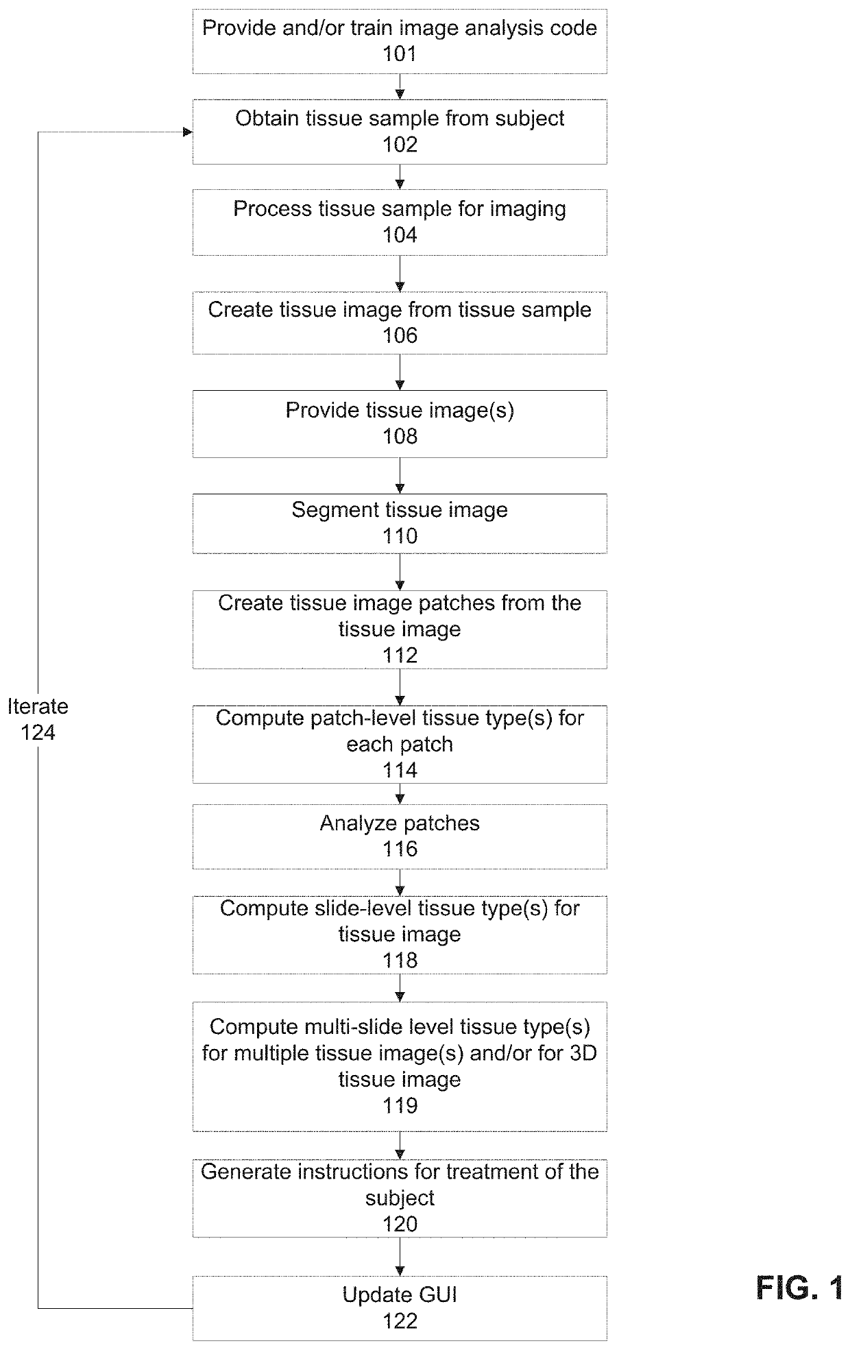
[0080]The present invention, in some embodiments thereof, relates to image processing and, more specifically, but not exclusively, to systems and methods for classification of pathological images.

[0081]As used herein, the term tissue, for example, as used with reference to tissue image, tissue sample, and tissue extracted from a subject, refers to a physically connected group of cells such as histological sample, for example, as obtained by a biopsy of an organ such as thyroid, and / or refers to individual cells or small clumps of cells such as a cytology sample, obtained from for example, a sample of fluid such as urine, cerebrospinal fluid, and / or scrapping of tissue suspended within an artificial fluid.

[0082]As used herein, the term slide-level tissue type may refer, for example, to a diagnosis made for the patient according to the tissue image created from the tissue sample extracted from the patient (optionally arranged on a slide). The diagnosis may be, for example, according t...


PUM


Abstract

There is provided a method of computing at least one slide-level tissue type for a tissue image of tissue extracted from a patient, comprising: receiving a tissue image of a slide including tissue extracted from the patient, segmenting tissue objects of the tissue image, creating a tissue image patches from the segmented tissue objects of the tissue image, classifying, by a patch-level classifier, each of the plurality of tissue image patches into at least one patch-level tissue type, wherein each of the classified tissue image patches is associated with a relative location within the tissue image, analyzing, by a slide-level analysis code, the classified at least one patch-level tissue type and associated relative location for each of the plurality of tissue image patches outputted by the patch-level classifier, for computing at least one slide-level tissue type for the tissue image, and providing the at least one slide-level tissue type.

Description

RELATED APPLICATIONS[0001]This application is a continuation of U.S. patent application Ser. No. 16 / 635,206 filed on Jan. 30, 2020, which is a National Phase of PCT Patent Application No. PCT / IL2018 / 050862 having International filing date of Aug. 2, 2018, which claims the benefit of priority under 35 USC § 119(e) of U.S. Provisional Patent Application No. 62 / 540,652 filed on Aug. 3, 2017. The contents of the above applications are all incorporated by reference as if fully set forth herein in their entirety.FIELD AND BACKGROUND OF THE INVENTION[0002]The present invention, in some embodiments thereof, relates to image processing and, more specifically, but not exclusively, to systems and methods for classification of images including tissue samples.[0003]A pathology report produced after reviewing a patient's biological tissue samples is often the gold standard in the diagnosis of many diseases. Most pathologist reviewed samples using a traditional microscope. Digital pathology is the...

Claims


Application Information

Patent Timeline
Patent Type & Authority Applications(United States)
IPC IPC(8): G06T7/00G06T7/11G16H10/40G16H50/20G06K9/46G06K9/62G06N3/08G06V10/50
CPCG06T7/0012G06T7/11G16H10/40G16H15/00G06K9/4642G06K9/6277G06N3/08G16H50/20G06T7/143G06T7/194G06V20/69G06V10/50G06T2200/24G06T2207/20021G06T2207/20076G06T2207/20081G06T2207/20084G06T2207/30024G06F18/2415
Inventor VEIDMAN, AVICHOREV, LOTAN
Owner NUCLAI LTD
Why Eureka
Try Eureka