
Method for analyzing lesion based on medical image

a technology of medical image and analysis method, applied in the field of medical image processing, can solve the problems of inability to generate and process, and the technology in the related art cannot show a performance which meets the purpose of lesion detection and evaluation

Pending Publication Date: 2022-06-23
VUNO INC
View PDF0 Cites 0 Cited by
  • Summary
  • Abstract
  • Description
  • Claims
  • Application Information

AI Technical Summary

Benefits of technology

The patent provides several improvements to existing methods for detecting and evaluating lesions in medical images. These improvements can help improve the process of diagnosing specific diseases.

Problems solved by technology

The inventors of the present disclosure have identified that some of technologies developed in the related art merely focuses on specifying the lesion which exists in the medical image, and is not capable of generating and processing, and providing information on the lesion in a form suitable for diagnosis of the specific disease.
That is, there is a situation in which in terms of providing required information suitable for the diagnosis of the specific disease, the technologies in the related art cannot show a performance which meets a purpose of lesion detection and evaluation.

Method used


Image

  • Method for analyzing lesion based on medical image
  • Method for analyzing lesion based on medical image
  • Method for analyzing lesion based on medical image

Examples


Embodiment Construction

[0040]Hereinafter, various embodiments are described with reference to the drawings. In the present specification, various descriptions are presented for understanding the present disclosure. However, it is obvious that the embodiments may be carried out even without a particular description.

[0041]Terms, “component,”“module,”“system,” and the like used in the present specification indicate a computer-related entity, hardware, firmware, software, a combination of software and hardware, or execution of software. For example, a component may be a procedure executed in a processor, a processor, an object, an execution thread, a program, and / or a computer, but is not limited thereto. For example, both an application executed in a computing device and the computing device may be components. One or more components may reside within a processor and / or an execution thread. One component may be localized within one computer. One component may be distributed between two or more computers. Furt...


PUM


Abstract

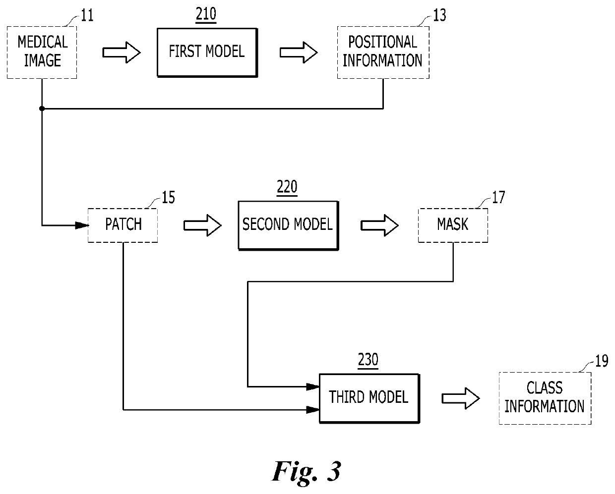
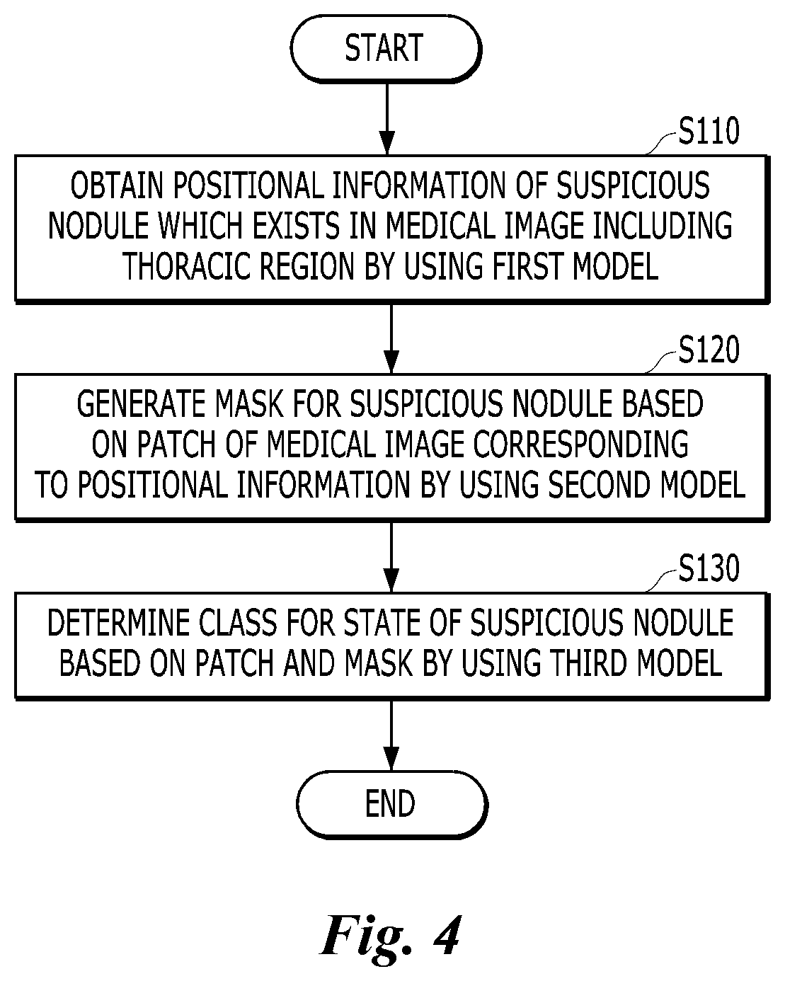
Disclosed is a method for analyzing a lesion based on a medical image, which is performed by a computing device. The method may include: obtaining positional information of a suspicious nodule which exists in the medical image; generating a mask for the suspicious nodule based on a patch of the medical image corresponding to the positional information; and determining a class for a state of the suspicious nodule based on the patch of the medical image and the mask for the suspicious nodule.

Description

CROSS-REFERENCE TO RELATED APPLICATIONS[0001]This application claims priority to and the benefit of Korean Patent Application No. 10-2020-0181634 filed in the Korean Intellectual Property Office on Dec. 23, 2020, the entire contents of which are incorporated herein by reference.BACKGROUNDTechnical Field[0002]The present disclosure relates to a method for processing a medical image, and more particularly, to a method for detecting and evaluating a lesion for a specific disease which exists in a medical image using artificial intelligence.Description of the Related Art[0003]A medical image is a material that allows physical states of various organs of the human body to be understood. The medical mage includes a digital radiographic image (X-ray), a compute tomography (CT), or magnetic resonance imaging (MRI).[0004]Research and technology development for an automation method for detecting a lesion of a specific disease is steadily continued based on the medical image.[0005]U.S. Pat. No...

Claims


Application Information

Patent Timeline
IPC IPC(8): G06T7/00G06T7/70G06T7/11G06V10/25G06T7/62G06T7/30G06V10/764G16H30/40G16H50/20
CPCG06T7/0016G06T7/70G06T7/11G06V10/25G06T7/62G06T7/30G06T2207/20081G16H30/40G16H50/20G06T2207/30064G06V2201/032G06T2200/24G06T2207/20084G06V10/764G06V10/82G06T7/0012G16H50/30G16H50/70G06T7/74G16H30/20A61B6/5217A61B5/055A61B8/5223G06N20/00G06T5/75
Inventor PARK, HYUNHOPARK, GWANGBEENLEE, SEUNGHO
Owner VUNO INC
Learn More