
Multiscale brain electrode devices and methods for using the multiscale brain electrodes

a brain electrode and multi-scale technology, applied in the field of multi-scale electrodes, can solve the problems of demonstrating the current limitation of epilepsy surgery, significant neurological deficit, and rare fast ripples

Pending Publication Date: 2022-07-07
MAYO FOUND FOR MEDICAL EDUCATION & RES
View PDF3 Cites 0 Cited by
  • Summary
  • Abstract
  • Description
  • Claims
  • Application Information

AI Technical Summary

Benefits of technology

The patent describes a new type of brain electrode that can be used to treat epilepsy. These electrodes are special because they have tiny parts embedded within larger ones. This allows for both stimulation and recording of brain activity. The benefits include the ability to detect and map the areas of the brain that are responsible for seizures, potentially preventing them from happening. The electrodes can also be used to deliver targeted therapy to treat seizures and can help to improve brain stimulation therapy for epilepsy.

Problems solved by technology

Unfortunately, the EZ does not currently have an apriori electrophysiological definition, and is only a concept acknowledging that resection of the SOZ does not always lead to seizure freedom.
However, many patients do not have an MRI lesion, or the lesion extends into functionally eloquent brain, e.g. brain regions supporting language, that cannot be removed without causing a significant neurological deficit.
Unfortunately, these patients make up 20-30% of pre-surgical evaluations at major epilepsy centers.
For patients with normal MRI scans only 30-50% of those finally deemed surgical candidates will achieve seizure freedom, demonstrating the current limitation of epilepsy surgery.
However, fast ripples were rarely recorded from the clinical macroelectrodes.
By the time a seizure is detected using conventional macroelectrodes, a large mass of neural tissue may have been recruited and the seizure can be difficult to abort.
In end vessel venules the MRES may disrupt the vessel wall and occlude the flow of blood.
The prolonged immobility, and subcutaneous tunneling of electrodes required by standard procedures result in significant post-operative pain and recovery delays in many patients.
Similarly, standard subdural and depth electrodes for used in the evaluation of intractable epilepsy are associate with increased risks of infection.

Method used


Image

  • Multiscale brain electrode devices and methods for using the multiscale brain electrodes
  • Multiscale brain electrode devices and methods for using the multiscale brain electrodes
  • Multiscale brain electrode devices and methods for using the multiscale brain electrodes

Examples


working example # 1 (

Working Example #1 (Chronic Subthreshold Cortical Stimulation to Treat Focal Epilepsy)

[0063]INTRODUCTION: Approximately one to three in 1,000 people have focal drug resistant epilepsy (DRE). Epilepsy surgery is the most effective treatment but may not be feasible when seizures originate from critical cortical areas, i.e. eloquent cortex. Despite evidence for efficacy, current approaches to focal brain stimulation rarely yield seizure free outcomes. In this working example, 13 patients were treated with continuous subthreshold electrical cortical stimulation, which led to suppression of interictal epileptiform discharges (IEDs) and improvement in clinical seizures.

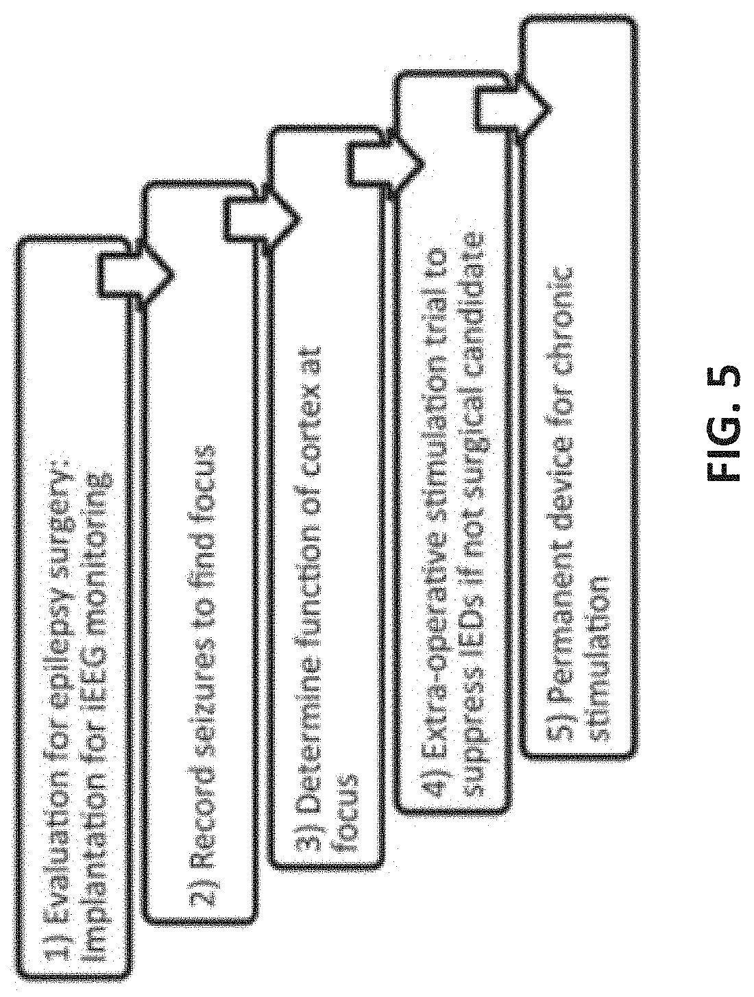
[0064]METHODS: Thirteen patients with focal DRE were deemed unsuitable for resective surgery following intracranial electroencephalography (iEEG) monitoring with surgically implanted subdural grid and depth electrodes (see FIG. 5 showing steps of patient evaluation and treatment). Pre-stimulation monitoring was typically se...

working example # 2 (

Working Example #2 (Multimodal Brain Mapping and Tracking)

[0068]INTRODUCTION: Multimodal imaging of brain integrates complementary neurophysiological signals from multiple modalities at multiple scales to make classifications of tissue epileptogenicity. The modalities include, but are not limited to, SPECT, PET, MRI structure, and wide bandwidth multi-scale Electrophysiology. SPECT, PET, and multiscale continuous (days to week, from hundreds of channels, sampled at 32 kHz) local field potential (LFP) intracranial recordings from human subjects have been acquired using some embodiments of the multiscale brain electrodes described above.

[0069]This example in conjunction with the other disclosure provided herein encompasses at least the following innovative concepts:

[0070]1) A database of normal SPECT, PET, and LFP data from specific brain structures (Frontal, Temporal, sub-Temporal, medial temporal Parietal, Occipital) and specific functional regions (motor, sensory, language).

[0071]2...

working example # 3 (

Working Example #3 (Microseizure Detection and HFO Detection)

[0110]INTRODUCTION: This example includes methods for: 1) labeling microdomain epileptiform events called microseizures and HFO in large-scale EEG records, and 2) automated labeling brain tissue as epileptic and identifying the ictal onset zone (IOZ), i.e the brain regions generating spontaneous seizures, based on spatial maps of microseizure and HFO density.

[0111]High frequency oscillations (HFOs) are emerging as a promising biomarker of epileptogenic tissue. HFOs are spontaneous EEG transients with frequencies traditionally considered to range from 60 to 600 Hz with 4<=cycles<50 that stand out from the background as discrete electrographic events. HFOs are usually divided into subgroups of high γ (60-80 Hz), ripple (80-250) and fast ripple (250-600) bands (Bragin et al. 1999).

[0112]Video-EEG (v-EEG) monitoring has long been a cornerstone in the evaluation of patients with seizure disorders. Generally, the primary goal of...


PUM


Abstract

Multiscale brain electrodes can be used for spatiotemporal mapping, probing, and therapeutic modulation of the human brain. The applications for such functional mapping and electrical stimulation modulation span, for example, neurological and psychiatric diseases, and brain rehabilitation.

Description

CROSS-REFERENCE TO RELATED APPLICATIONS[0001]This application claims the benefit of U.S. Provisional Application Ser. No. 62 / 334,843, filed May 11, 2016. The disclosure of the prior application is considered part of (and is incorporated by reference in) the disclosure of this application.STATEMENT AS TO FEDERALLY SPONSORED RESEARCH[0002]This invention was made with government support under NS095495, NS092882, and NS078136 awarded by the National Institutes of Health. The government has certain rights in the invention.BACKGROUND1. Technical Field[0003]This document relates to multiscale electrodes for spatiotemporal mapping, probing, and therapeutic modulation of the human brain.2. Background Information[0004]To effectively deliver electrical stimulation to probe and record the response of normal and pathological brain at the scales of single neurons, neuronal assemblies, local field potentials, and macroscopic EEG, the electrode surface area must be such that current can be safely d...

Claims


Application Information

Patent Timeline
Patent Type & Authority Applications(United States)
IPC IPC(8): A61B5/291A61B5/00
CPCA61B5/291A61B5/0006A61B5/4064A61B5/055A61B5/6868A61B5/7264A61B5/4094A61B5/053A61B5/4836A61B5/7257A61B5/726A61B5/7282A61B2562/04G16H50/20
Inventor BRINKMANN, BENJAMIN H.STEAD, SQUIRE MATTHEWWORRELL, GREGORY
Owner MAYO FOUND FOR MEDICAL EDUCATION & RES
Learn More