Method and apparatus for ultrasound imaging with biplane instrument guidance

a technology of ultrasound imaging and guidance, applied in the field of medical devices, can solve the problems of inability to achieve the desired accuracy of structures/features within the body, inability to provide 2d (two-dimensional) images, and inability to use linear array transducer probes

Inactive Publication Date: 2001-07-17
DIASONICS ULTRASOUND
View PDF15 Cites 220 Cited by
  • Summary
  • Abstract
  • Description
  • Claims
  • Application Information

AI Technical Summary

Problems solved by technology

Since such linear array transducer endocavitary probes provide one imaging plane at any time for viewing the interior of the body and / or another instrument (e.g., a biopsy needle) that may be guided into the body via the probe, the ultrasound images provided by such probes are generally limited to a single plane.
Unfortunately, such 2D (two-dimensional) images typically do not provide desired accuracy of structures / features within a body.
In applications where a relatively high level of imaging accuracy may be critical, such as biopsy needle guidance through or in proximity to sensitive bodily structures which may need to be avoided by the needle, such linear array transducer probes may not be practical.
Several techniques for improving the above-mentioned limitation(s) of single linear array transducer endocavitary probes have been proposed, but each is relatively limited as well.
However, end-fire multiplane single mechanical transducer probes cannot be used for biopsy guidance, since the biopsy needle is only visible in at most one plane of the rotation.
Moreover, side-fire multiplane single mechanical transducer probes are unable to provide viewing of a forward-penetrating biopsy need at all.
However, this type of biplane probe is limited to only one imaging plane, since the two imaging planes intersect over one transducer, not along the biopsy needle.
However, this second type of a biplane endocavitary probe does not allow viewing of a biopsy needle in either imaging plane.
In alternative embodiments, the ultrasound imaging probe may not provide a groove and / or other attachment features.

Method used


Image

  • Method and apparatus for ultrasound imaging with biplane instrument guidance
  • Method and apparatus for ultrasound imaging with biplane instrument guidance
  • Method and apparatus for ultrasound imaging with biplane instrument guidance

Examples


Embodiment Construction

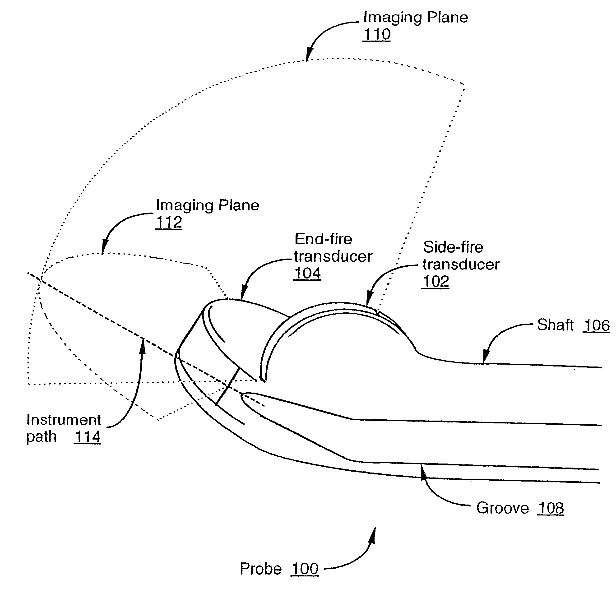
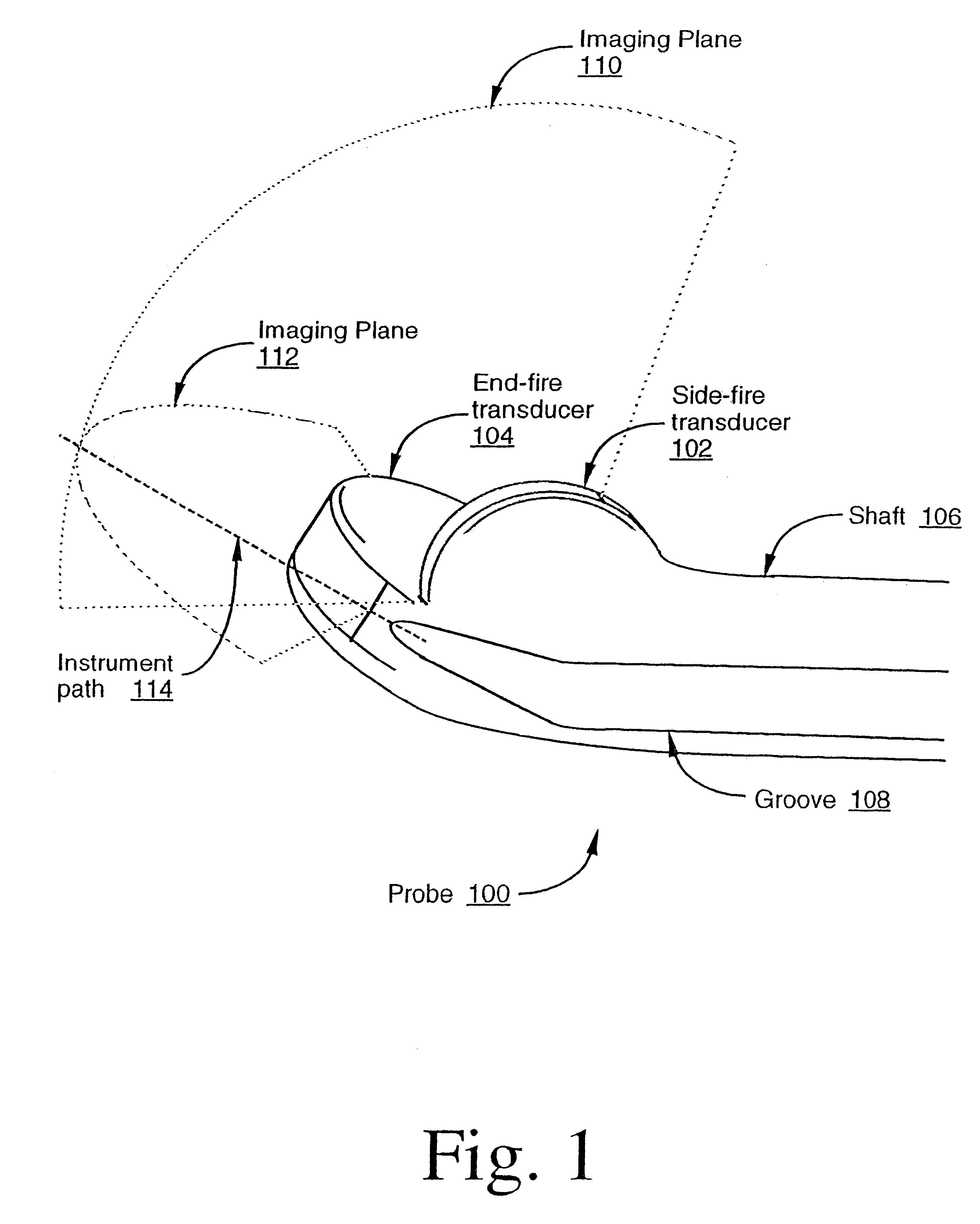
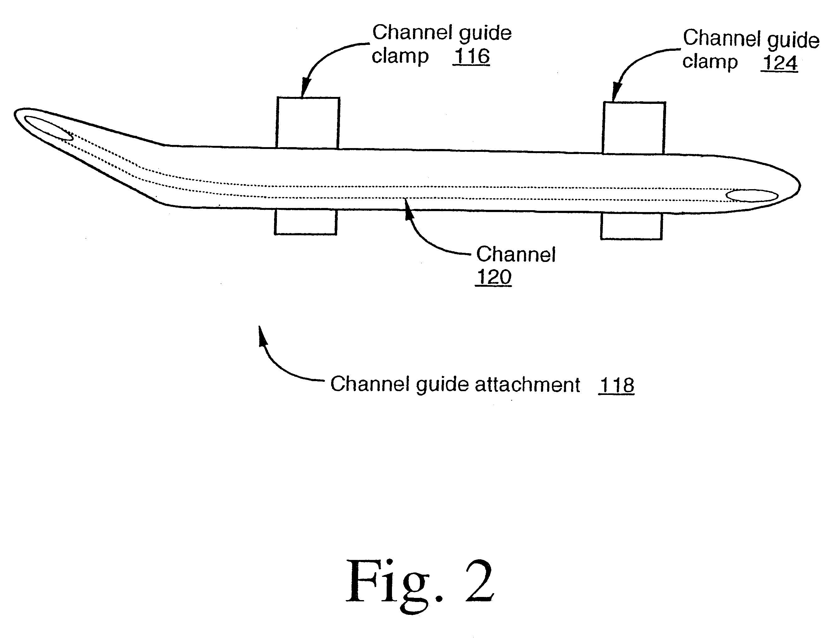
While the invention has been described in terms of several embodiments, those skilled in the art will recognize that the invention is not limited to the embodiments described. For example, while some types of transducers, ultrasound imaging device circuitry, probe / channel guide attachments, etc., have been shown and described, it will be appreciated that the invention is not limited to such. Accordingly, it will be appreciated that the invention may be embodied in various probe configurations and imaging system architectures that provide simultaneous viewing of an instrument (e.g., an endocavitary biopsy needle) in at least two ultrasound imaging planes.

Therefore, it should be understood that the method and apparatus of the invention can be practiced with modification and alteration within the spirit and scope of the appended claims. The description is thus to be regarded as illustrative instead of limiting on the invention.


PUM


Abstract

Methods and apparatuses for providing simultaneous viewing of an instrument in two ultrasound imaging planes. An ultrasound imaging probe is provided which can generate at least two ultrasound imaging planes. In one embodiment, the two imaging planes are not parallel (i.e., the planes intersect). An instrument path is positioned with respect to the planes such that an instrument may be simultaneously viewed in both imaging planes. In one embodiment, the instrument path is provided at an intersection that, at least partially, defines the intersection of the two imaging planes.

Description

1. Field of the InventionThe invention relates to the field of medical devices. More particularly, the invention relates to ultrasound endocavitary imaging devices.2. Background InformationVarious ultrasound techniques and devices have been developed for imaging the interior of a body (hereinafter, "body" refers to various types of subjects, such as humans, animals, non-living structures, etc.). One application of ultrasound imaging has been in the medical field, and in particular, in endocavitary probes (e.g., biopsy guidance endocavitary probes). Such probes may be used, for example, for endovaginal examination (e.g., to examine the uterus, ovaries, etc.), endorectal examination (e.g., to examine the rectal wall, prostate, etc.), and / or other medically-related applications. Typically, endocavitary probes include a linear array transducer positioned at the distal end of the probe that is to be inserted into a cavity of a body. The transducer provides an imaging plane for viewing st...

Claims


Application Information

Patent Timeline
Patent Type & Authority Patents(United States)
IPC IPC(8): A61B8/12A61B10/02
CPCA61B8/0833A61B8/0841A61B8/445A61B8/145A61B8/4488A61B8/12
Inventor LIN, GREGORY SHARAT
Owner DIASONICS ULTRASOUND
Why Eureka
Try Eureka