Versatile stereotactic device and methods of use

a stereotactic device and stereotactic technology, applied in the field of stereotactic devices, can solve the problems of poor image resolution in the scan slice direction (i.e., z-direction), heavy existing devices, and inability to perform "routine" office examinations, etc., and achieve accurate and reproducible results. , the effect of convenient us

Inactive Publication Date: 2004-01-27
THE BRIGHAM & WOMEN S HOSPITAL INC
View PDF36 Cites 122 Cited by
  • Summary
  • Abstract
  • Description
  • Claims
  • Application Information

AI Technical Summary

Benefits of technology

An exemplary stereotactic device and method of the present invention can thereby facilitate routine examinations of a patient or subject. In particular, the subject's head can be easily and repeatedly examined with the confidence that imaging scans taken at different time periods can be superimposed or compared directly. In some cases, portions of the patient's spine can also be viewed routinely, e.g., during regular, pre-and post-surgical planning examinations. The imaging modalities that can be used to advantage (or used with each other in any combination, except that the imaging modality that has a "fixed" --not adjustable--machine coordinate system should preferably be used to obtain the first, baseline, or initial set of imaging scans) include MR, CT, PET, SPECT, MEG, and other such imaging / radiologic scanning or spectroscopic techniques.

Problems solved by technology

Though there are a number of known devices and techniques for potential application to these types of examinations, these known devices and techniques are generally not suitable for "routine" office examinations, in which factors such as ease of use, speed of use, comfort, cost, accuracy and reproducibility are of major consideration.
Indeed, existing devices are often heavy, unwieldy, cumbersome and require that the devices be affixed to the subject using pins, screws, bolts, brackets, staples and the like.
A problem with this technique, however, is that the image resolution in the scan slice direction (i.e., the z-direction) is poor compared with the resolution in the scan in-plane (x-y direction).
Because of this poor resolution, it is difficult to make precise positional determinations.
Moreover, these methods also require a degree of anatomical knowledge which may strain the capabilities of the average MR technologist.
Clearly such methods are not suitable for "routine" office examinations.
Again, affixing the patient to the machine may makes the patient uncomfortable during the scan.
Stereotactic devices are typically fashioned from precision aluminum alloy and are very expensive for all except non-routine use.
Moreover, it is usually cumbersome and time consuming to affix these devices to the patient, adding to their unsuitability for routine examinations.
Though such devices can be used to determine the location of a head in x-y space, and to determine the relative location of each imaging "slice," they do not permit the position of a head to be fully determined, e.g., as where the head is tilted in the imaging plane.
In addition to the limitations described above, the prior techniques are not generally suitable for direct alignment of images obtained from different imaging modalities.
However, image processing has the drawback in that the resolution of the processed image is dependent on the quality of the scan data set.
Because of the difficulty in reproducing the machine coordinate system or because different machines are invariably associated with different, incompatible machine coordinate systems, it has not before been possible to relate directly scans from different imaging modalities.
Moreover, it is not always possible to align directly scans from the same imaging modality (e.g., MR imagers) when comparing images obtained from machines made by different manufacturers.

Method used


Image

  • Versatile stereotactic device and methods of use
  • Versatile stereotactic device and methods of use
  • Versatile stereotactic device and methods of use

Examples


Embodiment Construction

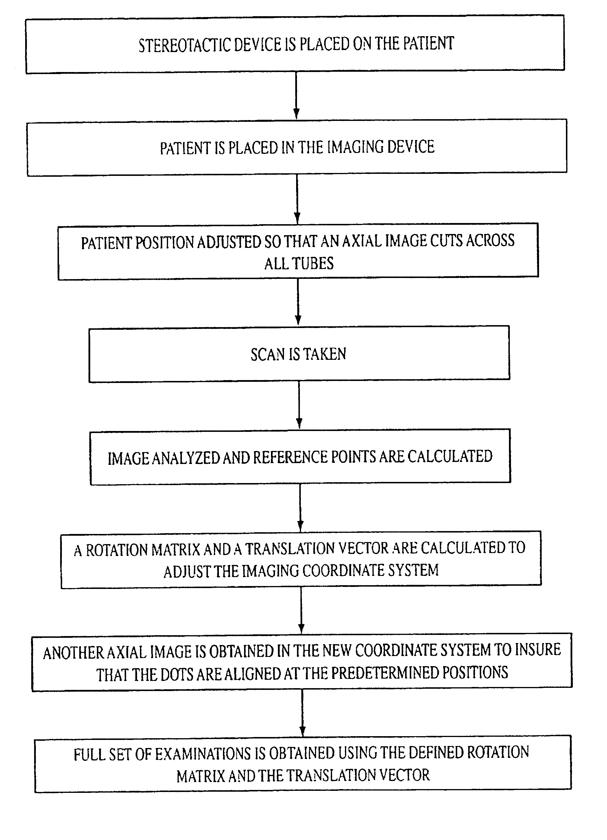
The illustrative device and method permit the alignment of routine scans to a pre-defined personal coordinate system. Once this alignment is performed, the location, orientation and size of, e.g., lesions can be reproducibly and precisely determined. It thus becomes possible to compare locations and size from different examinations at different times and sites using different machines for a given modality. It is further possible using the stereotactic device of the invention to provide for imaging scans that can be aligned directly and compared across different imaging modalities.

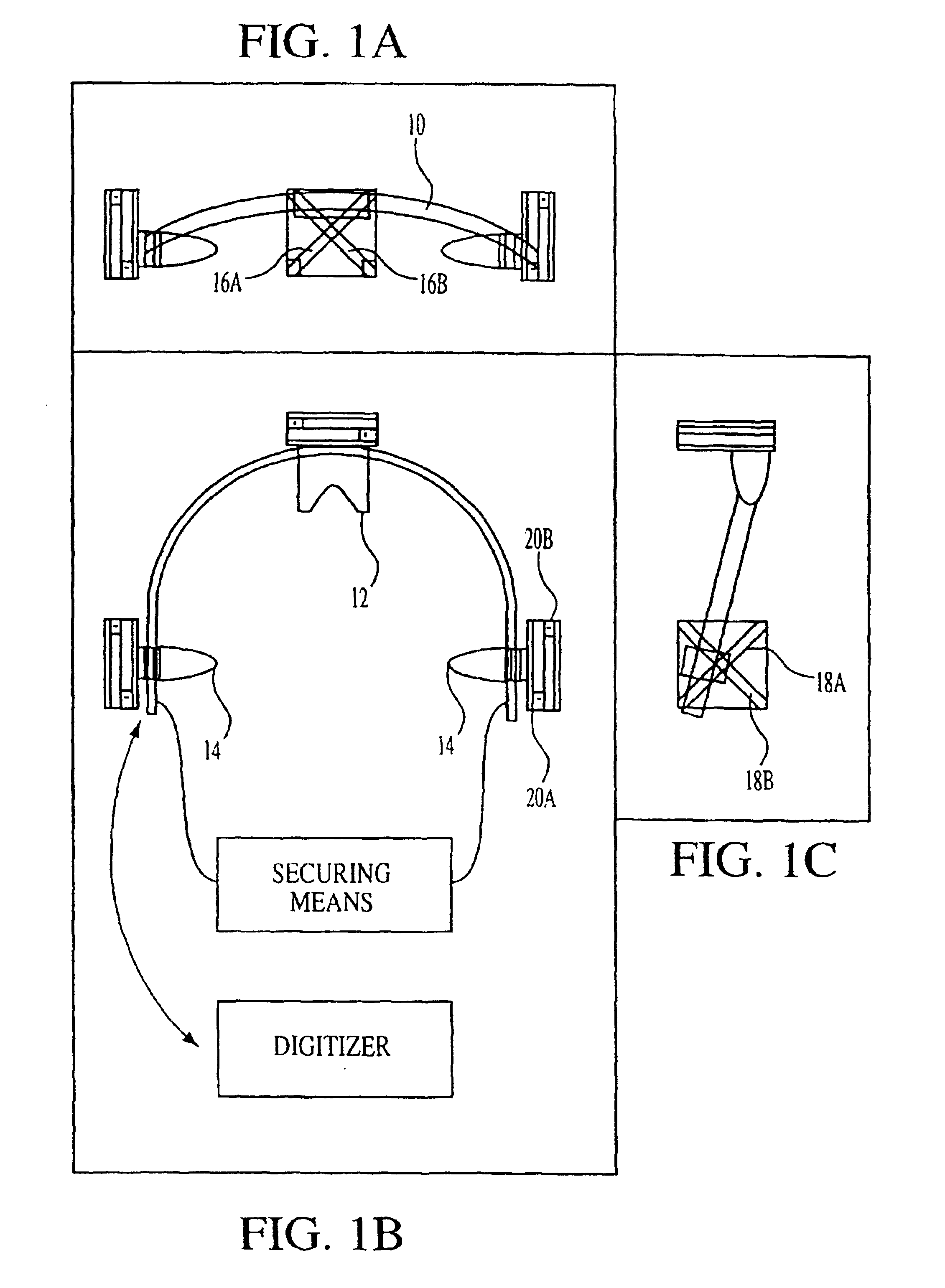
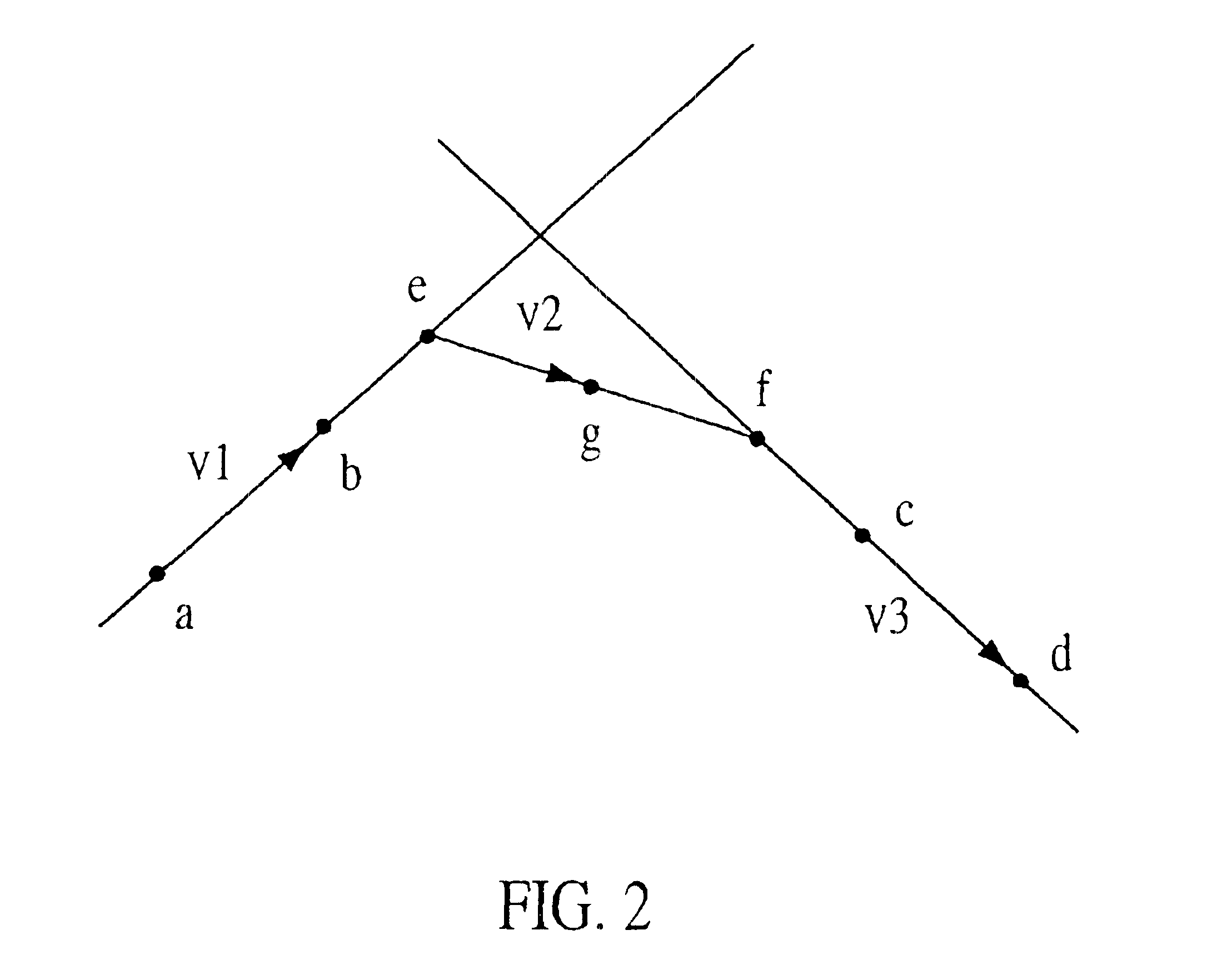
To accomplish further the objectives of the present invention, the following detailed description is provided which is directed to the lightweight frame, including non-invasive affixing means, localizing means and reference elements for defining a personal coordinate system and a computational procedure for the manipulation and / or alignment of coordinate systems to permit imaging scans to be taken at differ...


PUM


Abstract

A stereotactic device for use with an imager such as a magnetic resonance imager is disclosed, which permits an imaging scan to be taken with reference to a personal coordinate system (or PCS) that is independent of a machine coordinate system (or MCS). Methods using the device to obtain imaging scans are described such that the imaging scans are superimposable even if taken at different time periods using the same or a different imager. The device comprises a frame that can be reproducibly positioned on a subject and which is equipped with non-invasive affixing means and localizing means that provide the PCS. The device and methods of the invention are particularly well suited for routine initial or follow-up examinations, pre-surgical planning and post-surgical evaluation. Markers on a stereotactic device can be tracked during an MRI scan to compensate for patient motion during the scan.

Description

1. FIELD OF THE INVENTIONThe invention pertains to a versatile stereotactic device useful in a number of methods, including numerous modes of medical imaging. More particularly, the device and methods of the present invention relate to a non-invasive stereotactic method of reproducibly imaging portions of a patient's body, such as the patient's head and portions of the patient's spine in the proximity of the head. Thus, imaging modalities, including magnetic resonance (MR) imaging, magnetic resonance spectroscopy, computer-aided tomography (CT), positron emission tomography (PET), single photon emission computed tomography (SPECT), electroencephalography (EEG) or magnetoencephalography (MEG) and the like can be used to monitor, diagnose, or detect pathologic conditions and to follow their development, progress, arrest, or remission. The device and methods of the invention are especially applicable to permitting more routine head examinations, pre-surgical planning and providing post...

Claims


Application Information

Patent Timeline
Patent Type & Authority Patents(United States)
IPC IPC(8): A61B19/00A61B17/00
CPCA61B19/203A61B19/52A61B19/50A61B19/54A61B2017/00694A61B2019/5236A61B2019/5289A61B2019/5454A61B90/36A61B2090/364A61B90/14A61B34/10A61B2090/374A61B90/39A61B2090/3954
Inventor OSHIO, KOICHIPANYCH, LAWRENCE P.GUTTMANN, CHARLES R. G.
Owner THE BRIGHAM & WOMEN S HOSPITAL INC
Why Eureka
Try Eureka