Angiography method and apparatus

a technology of angiography and angiography method, applied in the field of angiography, can solve the problems of affecting the interpretation of images, affecting the quality of life, and affecting the accuracy of angiographic images, so as to improve accuracy and speed, and improve accuracy. the effect of tracking speed and accuracy

Inactive Publication Date: 2005-01-11
MARCONI MEDICAL SYST +1
View PDF12 Cites 195 Cited by
  • Summary
  • Abstract
  • Description
  • Claims
  • Application Information

AI Technical Summary

Benefits of technology

One advantage of the present invention is that it is a global technique which overcomes segmentation failures often encountered at vessel disjoints and overlaps by localized techniques.
Another advantage of the present invention is that it is compatible with both white blood angiography and black blood angiography.
Another advantage of the present invention is that it provides rapid and accurate vessel boundary estimation using propagation of geometric contours. The propagating can advantageously incorporate the gray scale image information through fuzzy membership classification of image elements in the neighborhood of the contour.
Another advantage of the present inven...

Problems solved by technology

Catastrophic medical events such as heart attacks and strokes that result from underlying vascular problems are a leading cause of death in the United States.
Many Americans suffer from chronic vascular diseases which degrade quality of life.
The analysis of angiographic images by medical personnel is often hindered by image imperfections or intervening non-vascular structures (e.g., bone, organs, and the like...

Method used


Image

  • Angiography method and apparatus
  • Angiography method and apparatus
  • Angiography method and apparatus

Examples


Embodiment Construction

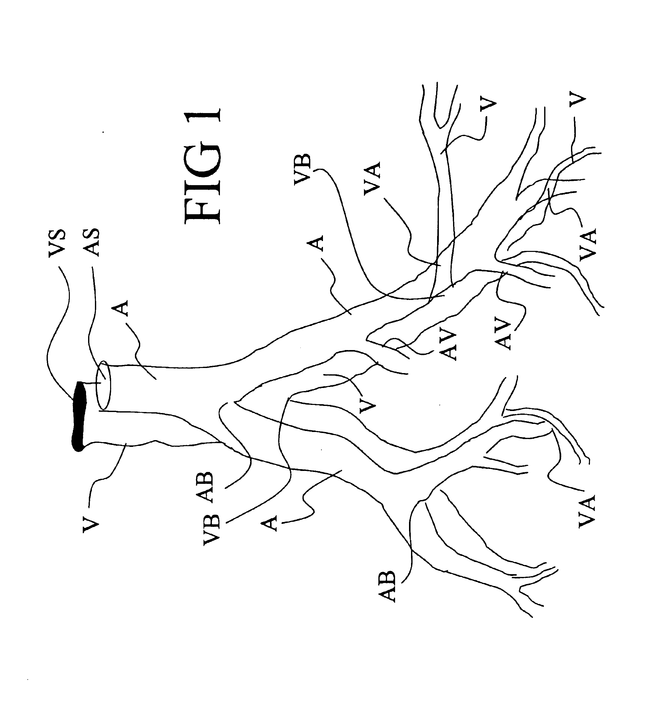
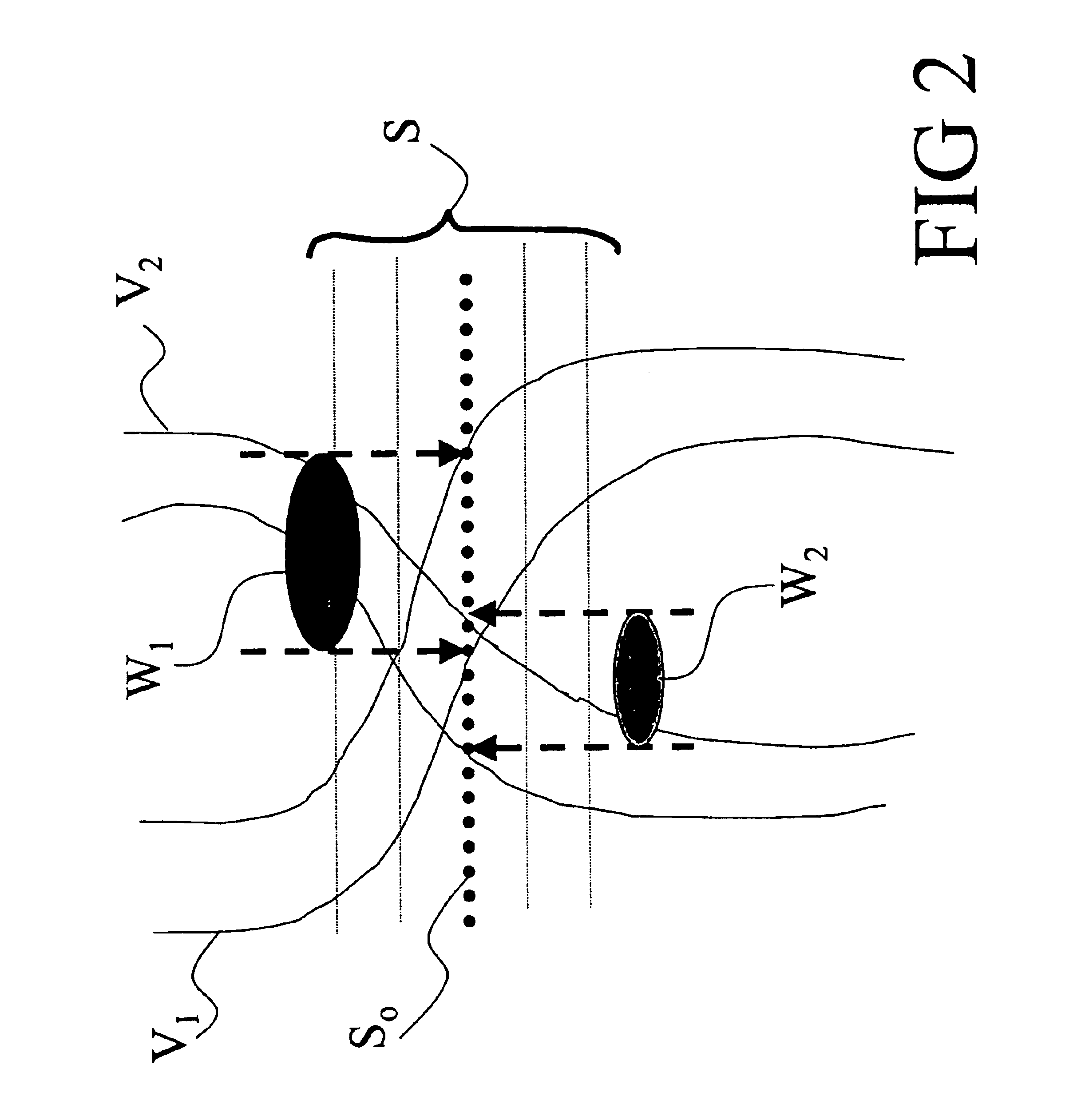
With reference to FIG. 4, a magnetic resonance imaging system that suitably practices angiographic imaging in accordance with an embodiment of the invention is described. Although the invention is described herein with respect to a magnetic resonance imaging embodiment, those skilled in the art will appreciate that the invention is applicable to a broad range of angiographic modalities and techniques, including but not limited to contrast-enhanced magnetic resonance angiography, non-contrast enhanced magnetic resonance angiography, computed tomographic angiography, and fused magnetic resonance / computed tomography angiographic techniques. The invention is also suitably practiced in conjunction with either white blood angiography (WBA) or black blood angiography (BBA).

With reference to FIG. 4, a magnetic resonance imaging (MRI) scanner 10 typically includes superconducting or resistive magnets 12 that create a substantially uniform, temporally constant main magnetic field B0 along a z...


PUM


Abstract

A two-dimensional slice formed of pixels (376) is extracted from the angiographic image (76) after enhancing the vessel edges by second order spatial differentiation (78). Imaged vascular structures in the slice are located (388) and flood-filled (384). The edges of the filled regions are iteratively eroded to identify vessel centers (402). The extracting, locating, flood-filling, and eroding is repeated (408) for a plurality of slices to generate a plurality of vessel centers (84) that are representative of the vascular system. A vessel center (88) is selected, and a corresponding vessel direction (92) and orthogonal plane (94) are found. The vessel boundaries (710) in the orthogonal plane (94) are identified by iteratively propagating (704) a closed geometric contour arranged about the vessel center (88). The selecting, finding, and estimating are repeated for the plurality of vessel centers (84). The estimated vessel boundaries (710) are interpolated (770) to form a vascular tree (780).

Description

BACKGROUND OF THE INVENTIONThe present invention relates to the medical imaging arts. It particularly relates to angiography using the magnetic resonance imaging (MRI) and computed tomography (CT) medical imaging techniques, and will be described with particular reference thereto. However, the invention will also find application in conjunction with other three-dimensional imaging modalities as well as in other imaging arts in which thin structures or networks with overlapping or furcated portions are advantageously differentiated from extraneous imaged structures and background noise or tracked in three dimensions.Catastrophic medical events such as heart attacks and strokes that result from underlying vascular problems are a leading cause of death in the United States. Plaque buildup on the inside of the vascular walls can lead to strokes, coronary heart disease, and other medical conditions such as vessel rupture. Many Americans suffer from chronic vascular diseases which degrade...

Claims


Application Information

Patent Timeline
IPC IPC(8): A61B6/00G06T5/00G06T7/00G06T7/60
CPCA61B6/481A61B6/504G06T7/0012G06T7/0083G06T7/0089G06T7/604G06T2207/30172G06T2207/10088G06T2207/20064G06T2207/20161G06T2207/30101G06T7/12G06T7/149G06T7/64
Inventor SURI, JASJIT S.LIU, KECHENGWU, DEE H.
Owner MARCONI MEDICAL SYST
Why Eureka
Try Eureka